
Clinical cases
Discover how clinicians around the world apply botiss biomaterials in daily practice. Browse our collection of clinical cases — from socket and ridge preservation to complex hard and soft tissue reconstruction — each documenting the materials used, the surgical approach and the clinical outcome.
-
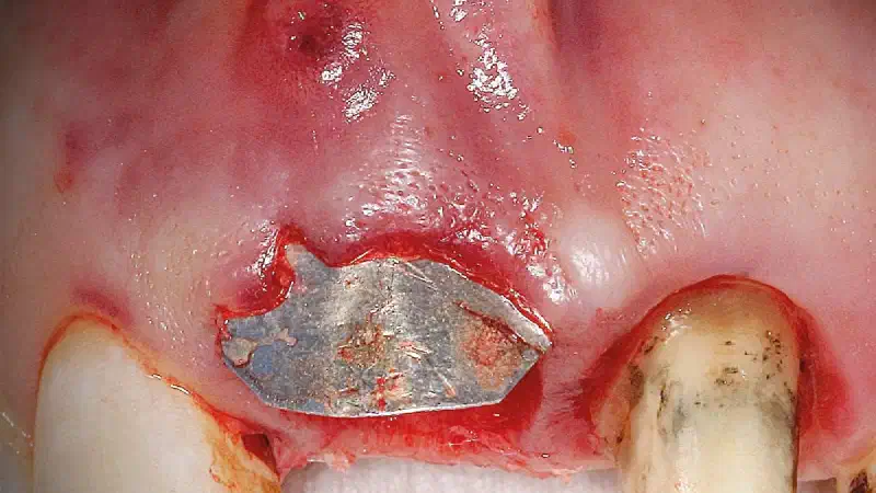
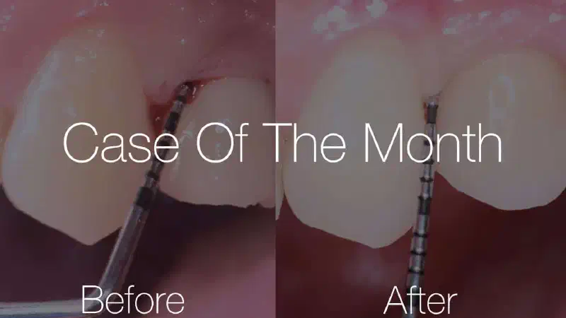
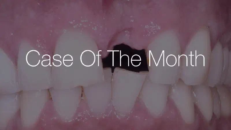
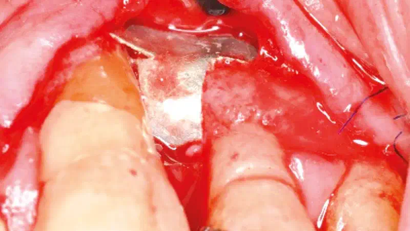
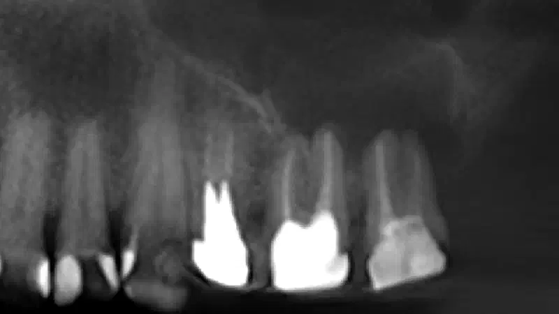
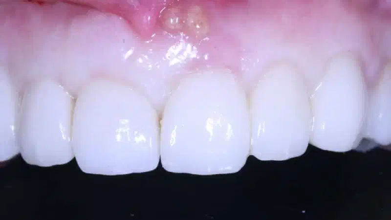
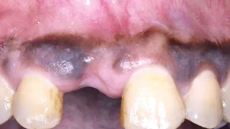
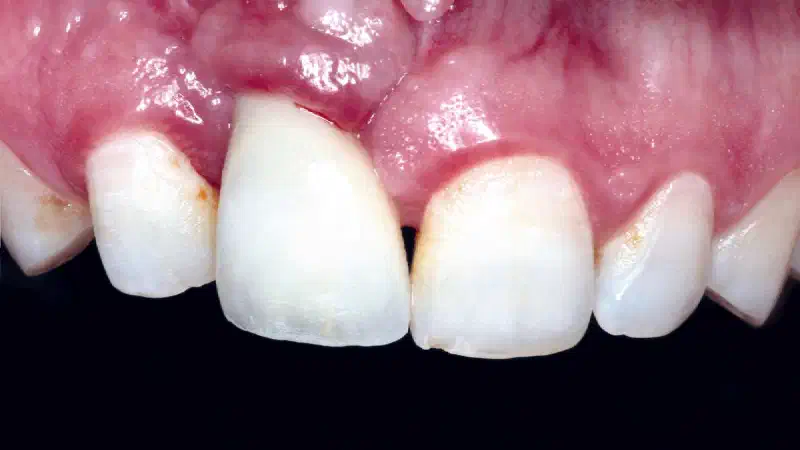
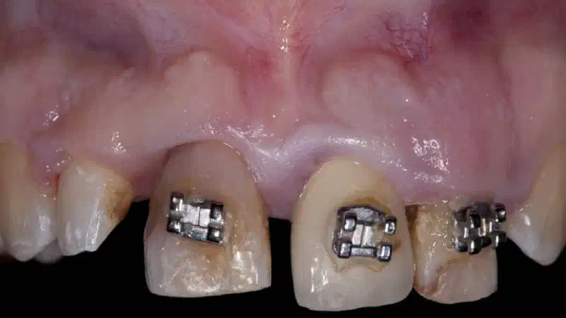
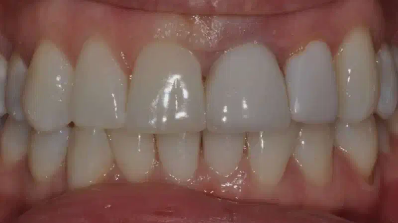
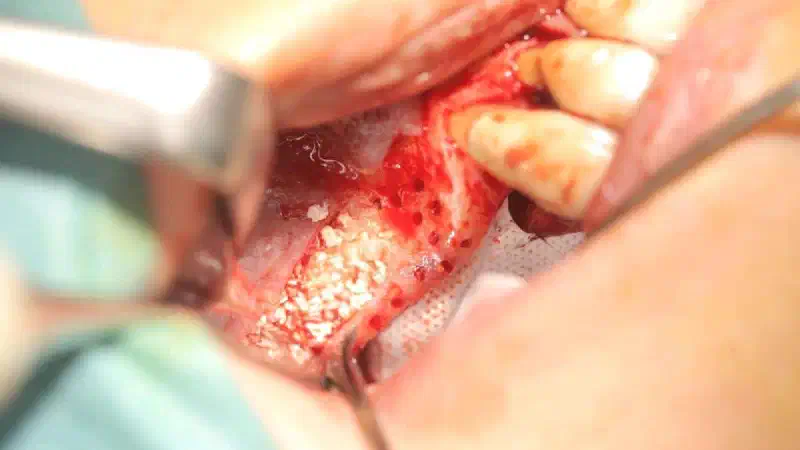
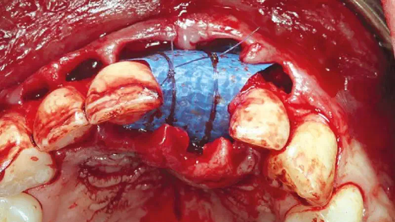
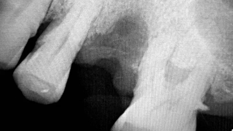
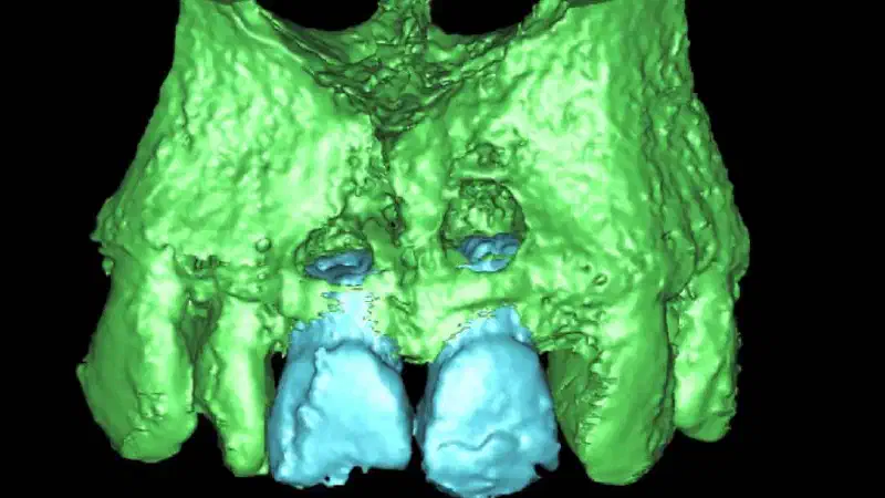
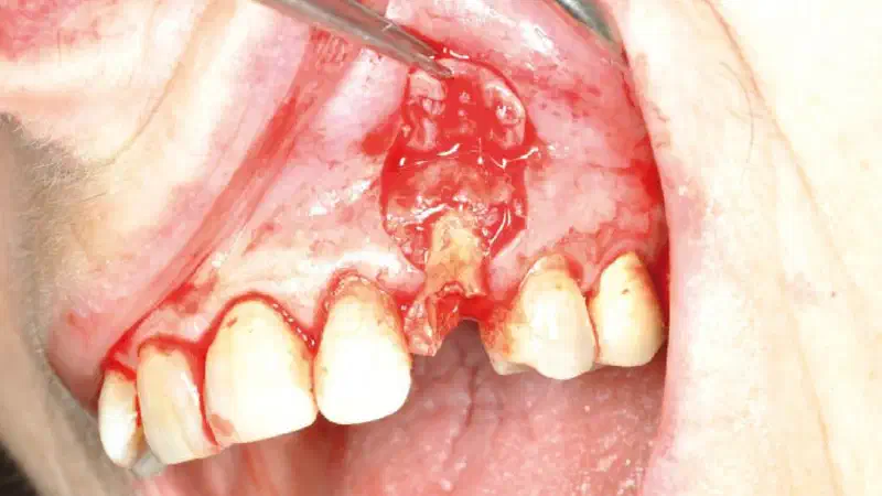
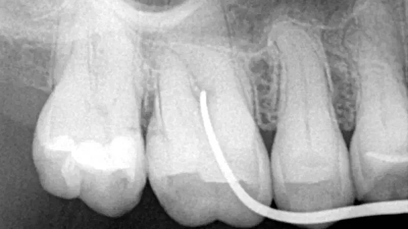
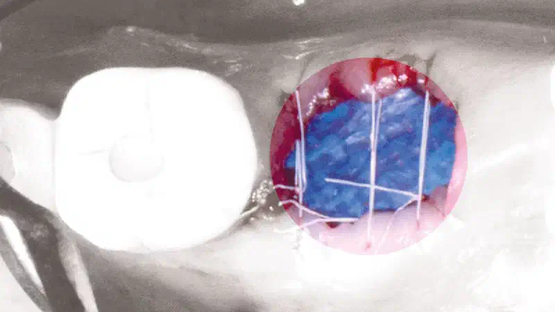
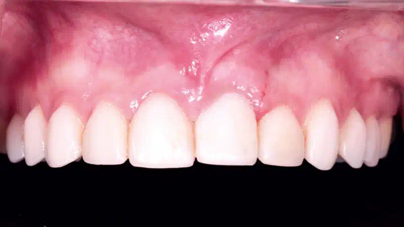
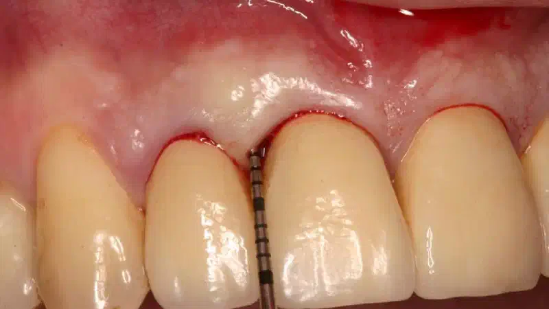
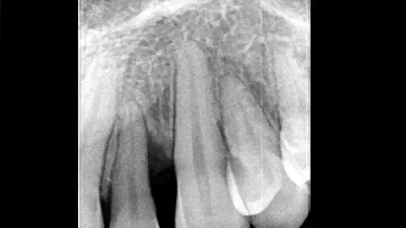
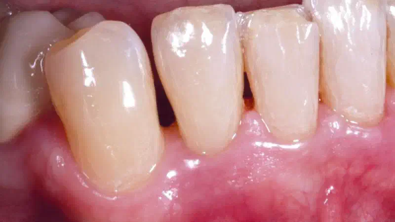
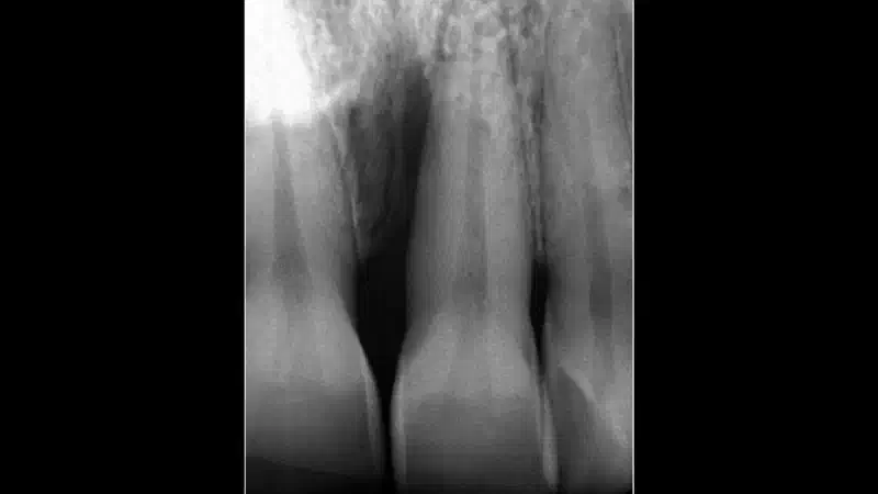
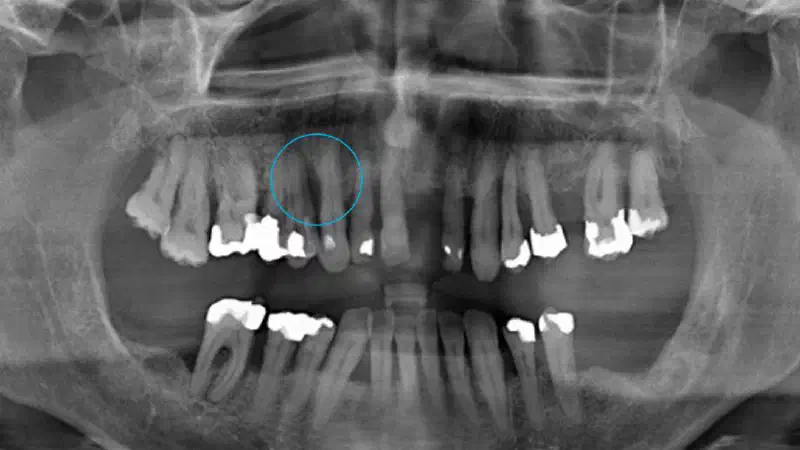
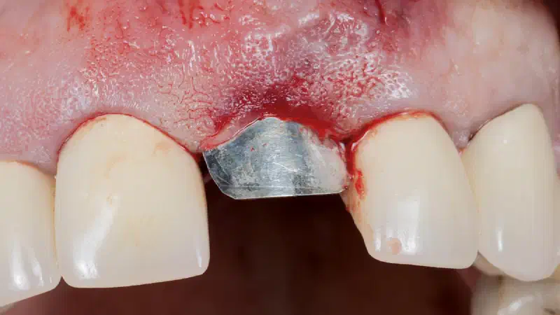
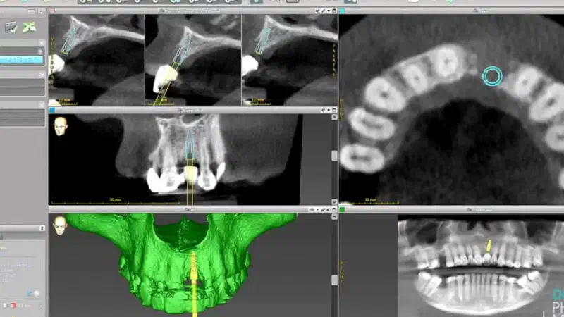
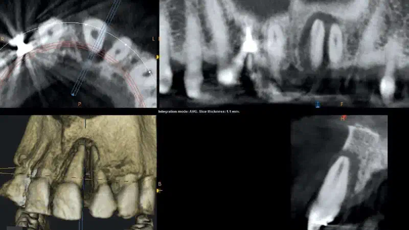
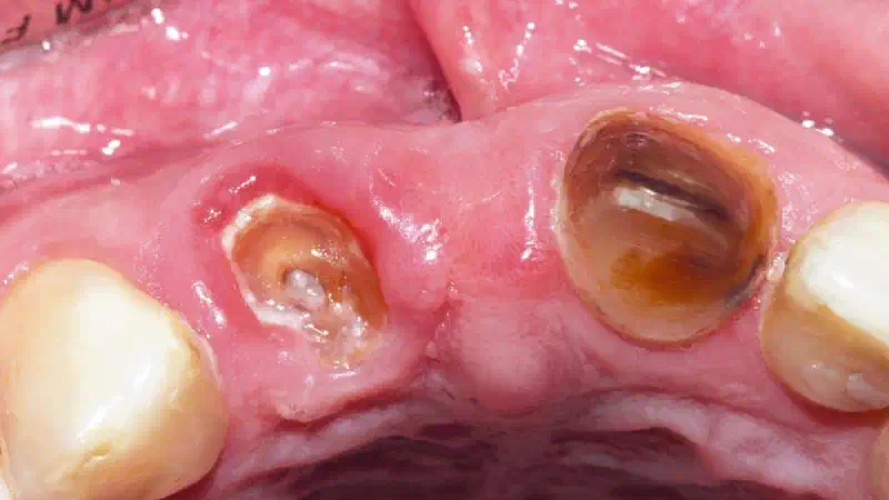
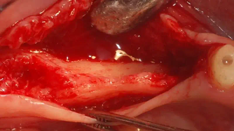
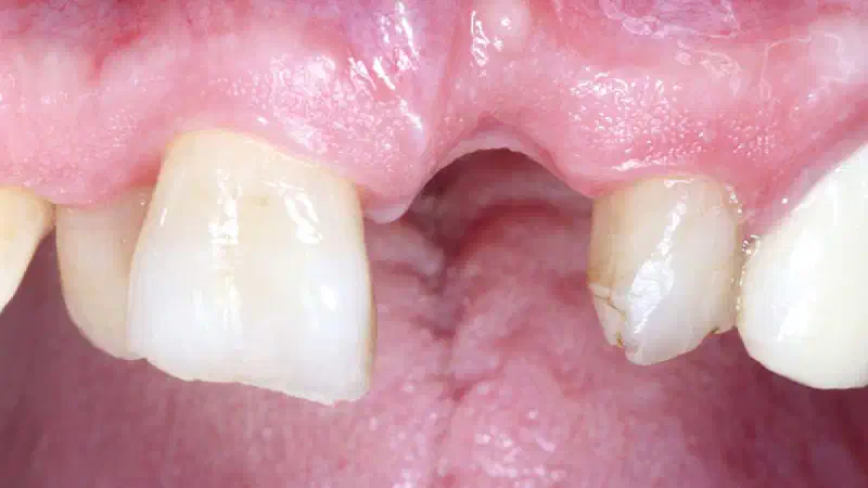
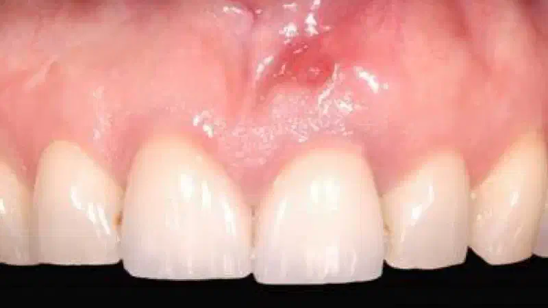
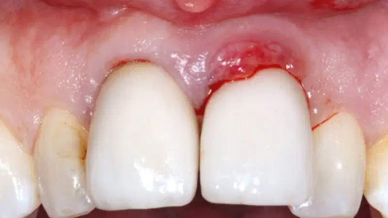
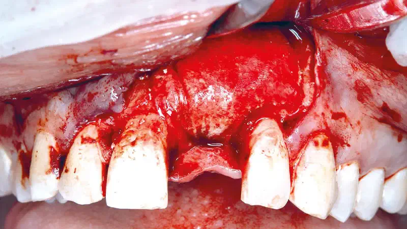
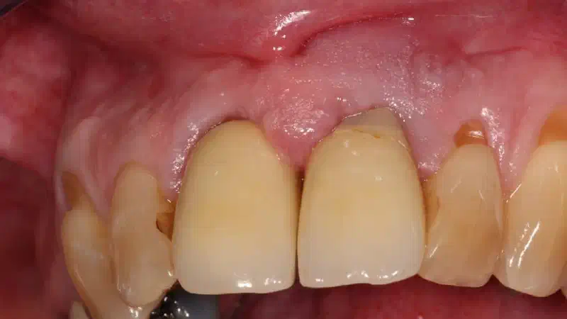
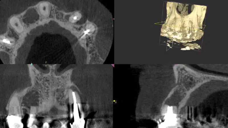
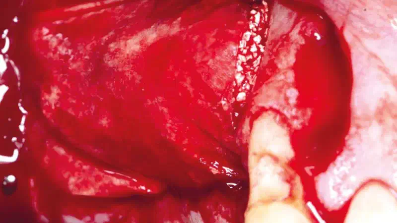
Aesthetic Buccal Flap and GBR for the Treatment of Extensive Apical Pathology Dr. Sebastián Bravo A 70-year-old patient presented with a large odontogenic cyst involving teeth #8 and #9 in the esthetic zone. Given the importance of preserving the natural dentition whenever biologically feasible, a conservative surgical approach was selected instead of extraction and implant placement. The primary clinical challenge was the management of an extensive periapical lesion while maintaining the integrity of the gingival architecture and avoiding esthetic complications such as scarring or soft tissue recession.
-
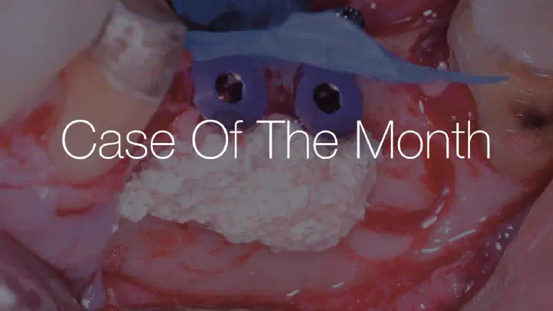
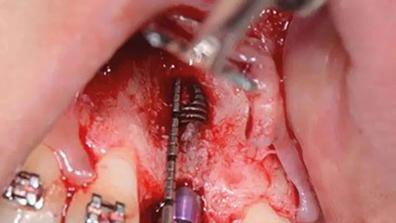
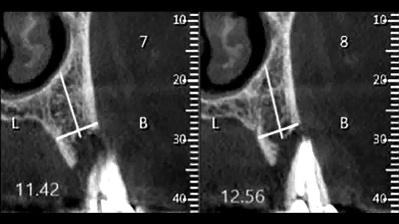
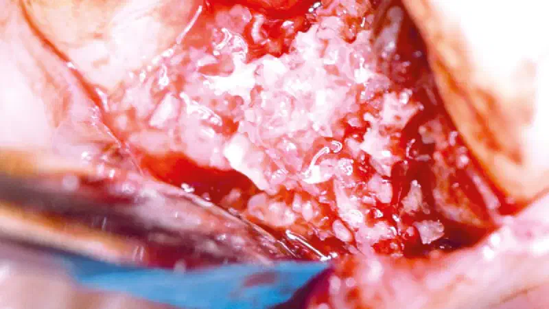
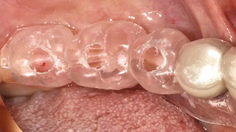
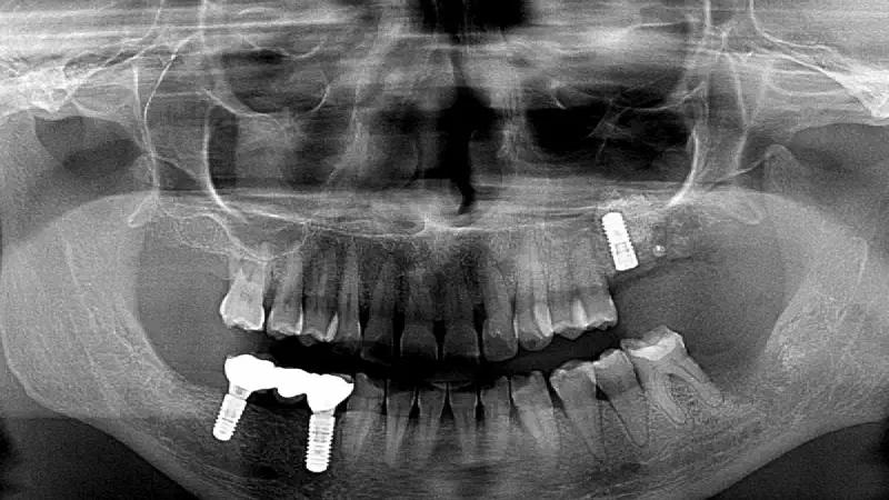
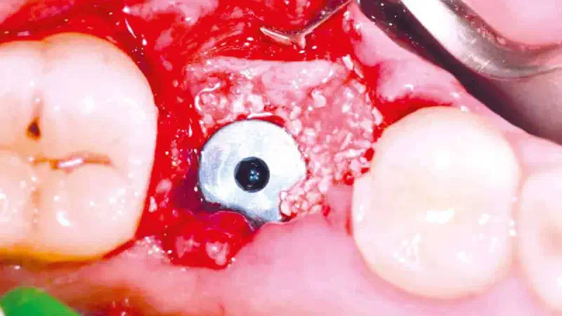
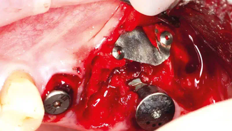
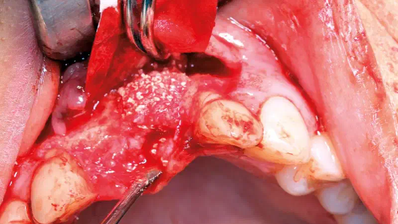
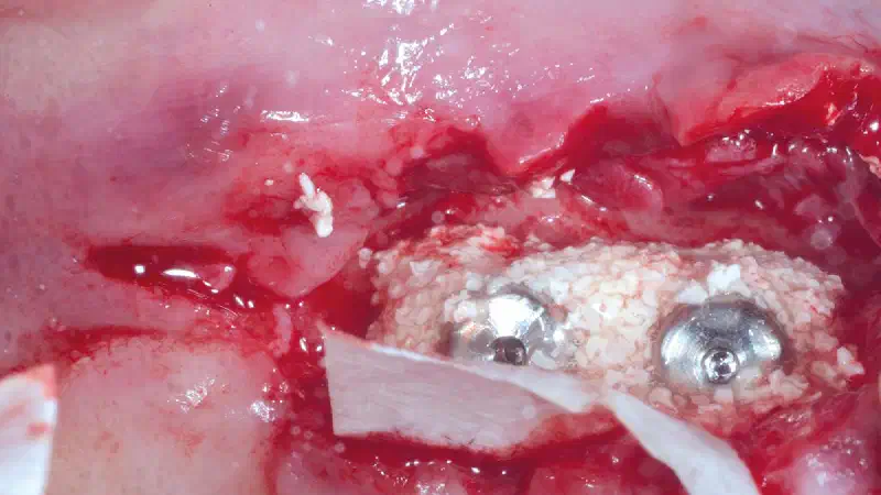
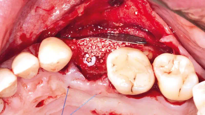
Sinus Augmentation and Lateral Ridge Reconstruction with Stable 3-Year Results Dr. Reto Morger A patient presented with insufficient vertical and horizontal bone volume in the posterior maxilla, requiring combined augmentation procedures to enable implant placement. Due to the reduced residual bone height and compromised ridge contour, a simultaneous approach consisting of sinus floor elevation, implant placement, and lateral ridge augmentation was performed. Following flap elevation, a lateral window technique was used to access the maxillary sinus. Careful elevation of the Schneiderian membrane was achieved without complications, creating sufficient space for graft placement. Implant insertion was performed simultaneously, achieving primary stability despite the limited native bone.
-
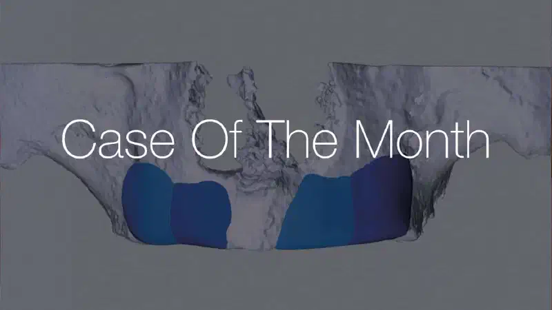
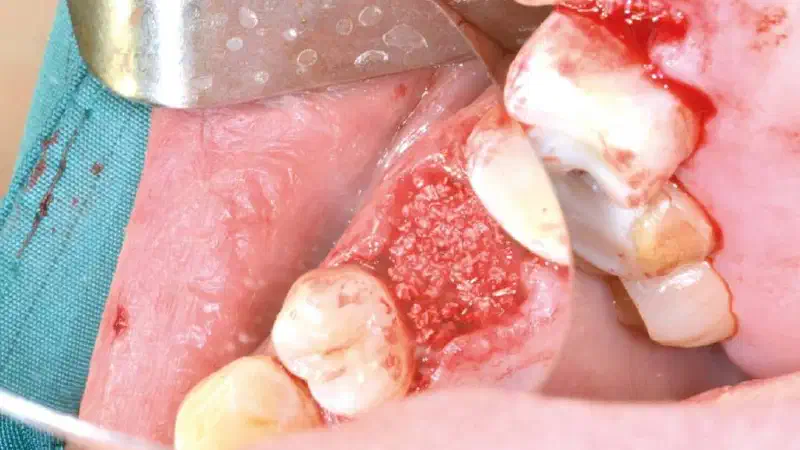
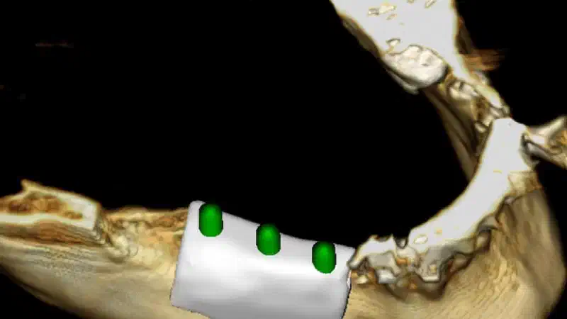
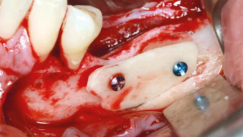
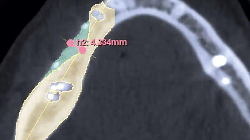
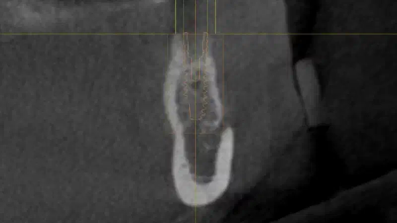
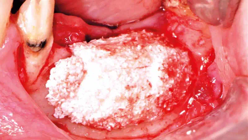
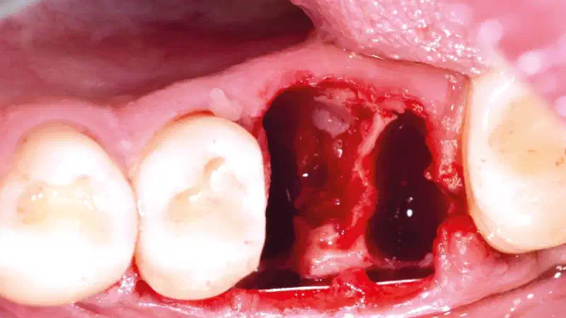
A 44-year-old female patient presented with missing teeth in the mandibular right posterior region (45–47). Clinical and CBCT evaluation revealed a severe horizontal ridge defect with limited bone width and reduced vertical dimension, making implant placement impossible. Residual bone width: approx. 2.4mm Residual bone height: 9.2–9.6 mm Indication: horizontal ridge augmentation prior to implant placement Surgical Procedure A full-thickness flap was elevated and the recipient site was prepared. Autologous bone was harvested from the retromolar region using a safe scraper.
-
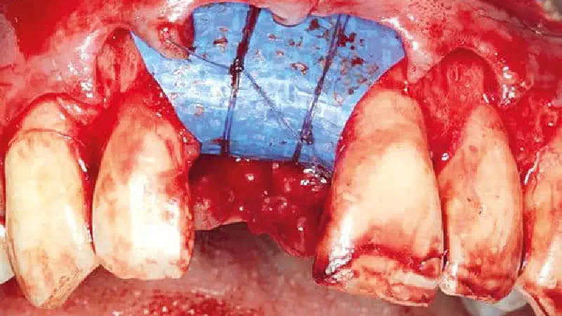
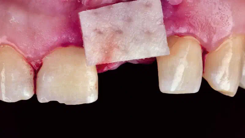
A patient presented with a failing maxillary anterior tooth in the aesthetic zone associated with a missing buccal bone wall, representing a high-risk situation for immediate implant therapy. An atraumatic extraction followed by immediate implant placement was performed. Due to the absence of the facial bone, the NOVAMag® SHIELD was applied as a rigid protective barrier in a flapless minimally invasive approach, allowing controlled buccal contour augmentation using cerabone® +HyA and preventing soft tissue collapse during healing. CTG from the palate was positioned and sutured in the pouch to enhance the buccal mucosal thickness.
-
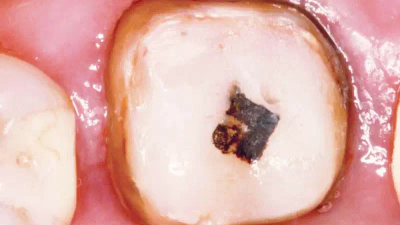
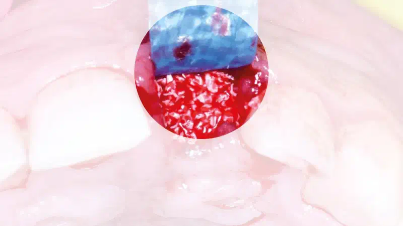
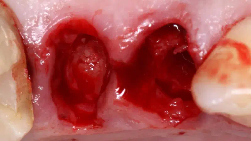
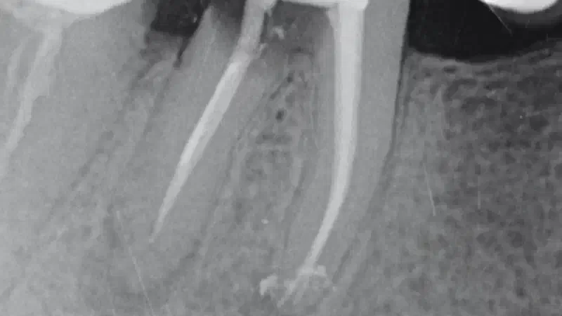
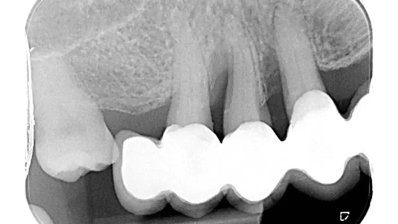
**Dr.**Alessandro Rossi The patient presented with a single-rooted tooth indicated for extraction. The Periodontal charting of the site showed a probing dept of 10 mm only on the palatal aspect, compatible with the diagnosis of vertical fracture. The periapical radiograph demonstrated deep root decay with associated hard tissue destruction. The tooth was removed atraumatically, and on the palatal aspect of the root was evidenced a vertical fracture. The extraction socket was subsequently thoroughly debrided to eliminate granulation tissue and ensure a clean recipient site. Intraoperative assessment revealed a buccal bone thickness of ≤ 1 mm. To preserve alveolar ridge dimensions and prepare the site for a staged implant approach, the socket was grafted with a bone substitute material for ridge preservation. For simultaneous soft tissue augmentation and socket sealing, mucoderm ® , was used in a combined onlay–interpositional technique. The buccal and palatal portions of the matrix were inserted beneath the respective full-thickness flaps, while the central portion was positioned over the crestal aspect of the socket and intentionally left exposed to the oral cavity to achieve socket sealing. Wound closure was accomplished with interrupted sutures placed interproximally and an internal horizontal mattress suture connecting the mid-facial and mid-palatal aspects of the flaps to ensure stable fixation and gentle compression of the matrix.
-
In this clinical case, a patient presented with a severely compromised upper right central incisor (tooth 1.1), characterized by significant probing depth, advanced bone loss and complete resorption of the buccal bone wall as confirmed by pre-operative CBCT. The tomographic images confirm an oblicual root fracture. The specific clinical situation suggested the planning of an atraumatic extraction followed by a ridge preservation and delayed implant placement in the anterior zone.
-
A 69-year-old female patient presented with a pronounced bucco-lingual ridge collapse and an insufficient band of keratinized mucosa. After bone augmentation with cerabone® +HyA , three implants were placed six months later. To enhance peri-implant soft tissue quality, a second-stage procedure was performed using mucoderm®, stabilized with a 7 mm NOVAMag® S fixation screw to ensure optimal adaptation and stability. Primary closure was achieved using reinforced e-PTFE sutures. At three months, clinical evaluation and STL imaging confirmed a wider band of keratinized mucosa, improved vestibular depth, and more than 1 mm of vertical and horizontal soft tissue gain.
-
Case presentation A 35-year-old systemically healthy female patient with a positive smoking history presented after completion of non-surgical anti-infective periodontal therapy. Despite regular supportive care at 3-month intervals, several sites exhibited persistent deep probing depths. The patient was motivated toward smoking reduction and enrolled in surgical regenerative treatment. As part of the comprehensive therapy plan, all four third molars were extracted to eliminate additional periodontal risk factors. Treatment Following debridement and root surface conditioning, guided tissue regeneration was performed using maxgraft® +HyA XS in combination with Emdogain® (Straumann Group). The surgical approach aimed at the reconstruction of intrabony defects and the re-establishment of periodontal attachment.
-
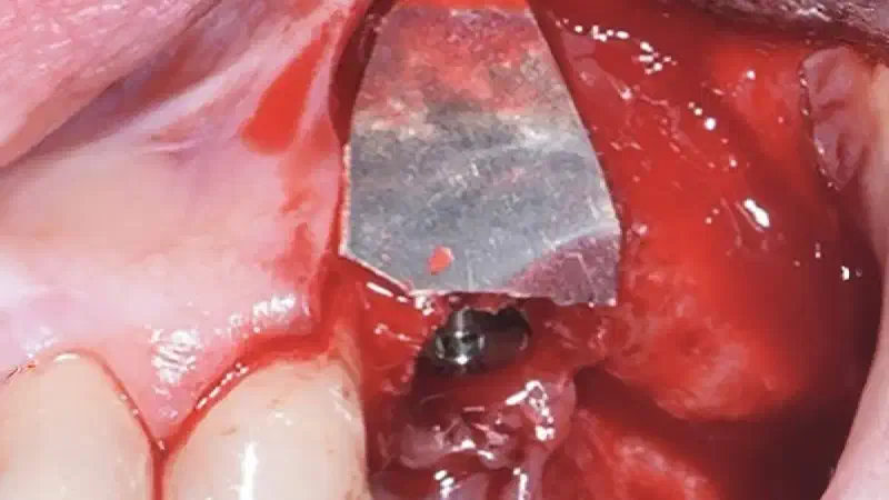
A patient presented with a peri-implant bone defect characterized by bone resorption compromising the stability and long-term prognosis of the implant. To re-establish sufficient bone volume and ensure implant longevity, a regenerative surgical intervention was performed using a guided bone regeneration (GBR) approach. The treatment featured cerabone® +HyA for grafting, complemented by the Shield Technique using fully resorbable magnesium-based NOVAMag® SHIELD to guide bone regeneration. By the 12-month follow-up, bone regeneration had advanced even further, and implant stability was significantly strengthened, confirming the procedure’s lasting success. A CBCT scan at 6 months revealed promising results, which were even more striking at the 12-month mark, demonstrating optimal bone integration and implant stability.
-
A patient presented with severe gingival recession and pronounced inflammation affecting tooth #24. Following extraction of the tooth and thorough debridement of the infected site, a complete loss of the vestibular wall and a slight resorption of the lingual wall were identified. In this situation, NOVAMag® membrane was cut to shape and given a suitable convexity and used as a rigid plate and fixated with two resorbable NOVAMag® fixation screws . The membrane was fixated of the lingual side only in this case. A narrow ⌀ 3.2 × 13 mm implant was placed. The remaining peri-implant gaps were filled with maxgraft® granules , and the augmentation site was finally covered with a collprotect® membrane .
-
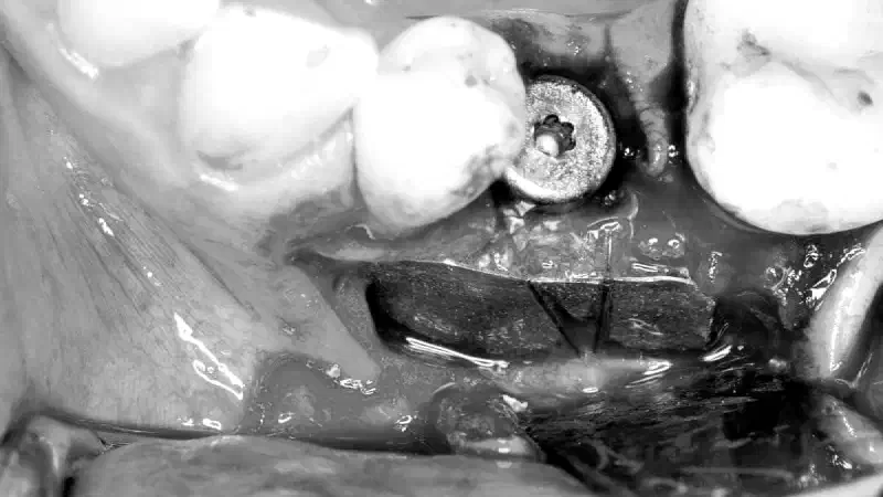
A young male patient presented with an inflamed lower right first molar (tooth 46). Due to the poor prognosis of the tooth, extraction was indicated and performed. Given the extent of local inflammation, no immediate implant placement was planned. Following a healing period, clinical and radiographic evaluation revealed insufficient volume and contour of the alveolar ridge, as anticipated. The mesial and distal bone walls remained intact, forming a classic four-walled defect. Autologous bone chips were harvested and mixed with cerabone® +HyA to create a cohesive grafting material. A NOVAMag® membrane was selected for this procedure due to its excellent space-maintaining properties and full resorbability. The membrane was folded over the augmented site and fixated both lingually and buccally with NOVAMag® fixation screws.
-
A healthy, 54-year-old female patient presented with peri-implantitis and aesthetic concern in the anterior maxilla. Discrepancy in gingival zenith and hard and soft tissue deficiencies were also evident. Following a non-surgical therapy to stabilize the soft tissue conditions, a reconstructive surgical approach was performed using mechanical and chemical implant surface decontamination followed by the filling of the intraosseous defect and simultaneous horizontal bone augmentation using maxgraft ® allogenic granules and collprotect ® membrane. In addition, a coronally advanced flap was performed on the adjacent teeth to improve the soft tissue conditions. Once the connection phase was completed, a provisional crown was placed during the healing period. Peri-implant parameters were monitored, which demonstrated stable peri-implant conditions after which the final restoration was delivered.
-
A 52-year-old male patient presented with severe swelling accompanied by purulent discharge from the anterior palate. Based on the clinical and radiographic findings, a preliminary clinical diagnosis of a radicular cyst associated with the maxillary incisors was established. The treatment plan was formulated to address both the pathological lesion and the resultant bony defect. The proposed treatment strategy involved a two-stage approach: initial marsupialization to reduce cyst size and promote bone formation, followed by cyst enucleation and GBR.
-
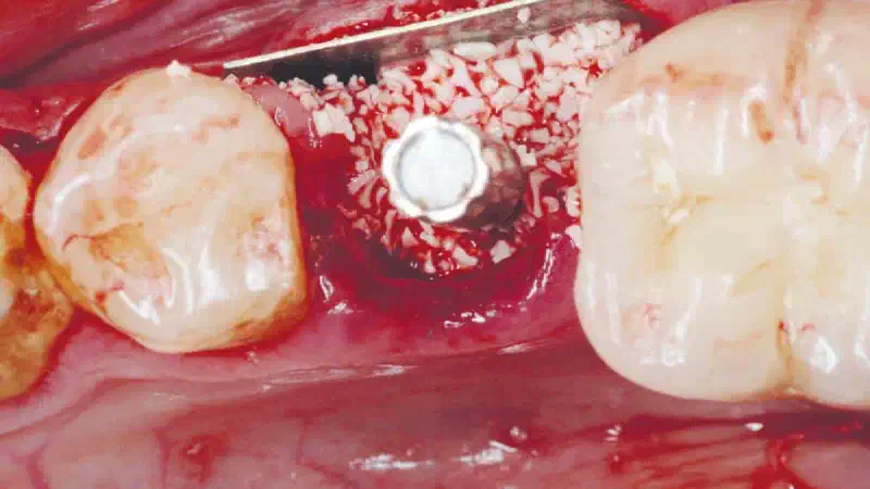
In the posterior mandible (lower right molar region), the edentulous site presented with insufficient hard and soft tissue volume. A two-stage Guided Bone Regeneration (GBR) surgery was performed to gain minor vertical height and major horizontal width, creating the necessary foundation for implant placement. Additional grafting for soft tissue thickness was performed with mucoderm® in same surgery. GBR was performed with permamem® membrane secured by NOVAMag® fixation screws (2 lingually, 2 buccally). Grafting material was 50% cerabone® +HyA and 50% autologous bone scraping placed on decorticalized site. After a healing period of six months, the augmented site exhibited adequate hard and soft tissue volume, allowing for successful implant placement with high primary stability.
-
Two-stage GBR in the aesthetic zone with maxgraft® +HyA cortico-cancellous Dr. Frank Kloss A Patient presented with a severe horizontal defect due to a missing central incisor. Guided bone regeneration was performed using the novel allogenic bone substitute material maxgraft® +HyA cortico-cancellous together with a long-lasting barrier membrane ( Jason® membrane ). The volume stability of the graft was confirmed by control CBCT at three months. Implant placement was performed after six months of healing. The one-year follow-up control demonstrates a stable clinical outcome.
-
Reconstruction of the anterior maxilla using the allogenic shell technique Dr. Pamela Ruiz A horizontal and vertical defect of upper central area was noticed due to trauma in a 53-year-old female patient. Steps planification include orthodontic treatment, horizontal and vertical bone regeneration with allogenic cortical plates ( maxgraft® cortico ) filled with a mix of autogenous and allogenic bone covered with Jason® membrane . After 9 months healing, reentry and installation of immediately loaded dental implants were performed and final restoration was made after 4 months. The 4-year follow up control demonstrates a stable clinical outcome.
-
Replacement of a failing maxillary lateral incisor in a 36year old female patient INITIAL SITUATION A 36-year old female patient in good general health presented with a history of a traumatic experience when 12 years old associated to the upper left lateral incisor tooth. The tooth subsequently became ankylosed, discoloured and a step deformity developed over time. The tooth was not root treated, and upon presentation was clearly being affected by internal resorption.
-
From Explantation to Functional Restoration: A Multi-Phase Maxillary Rehabilitation Case Dr. Luka Markovic A 60-year-old patient presented with complications arising from poorly positioned maxillary implants. The treatment plan included explantation, followed by guided bone regeneration (GBR) utilizing autologous bone, cerabone® +HyA , and the NOVAMag® membrane . Autologous bone chips were mixed with cerabone® +HyA to augment the site, while the NOVAMag® membrane was carefully shaped, positioned over the augmentation area, and secured with NOVAMag ® fixation screws .
-
Staged implant placement following horizontal GBR with cerabone® +HyA, autologous bone, permamem® & Jason® membrane in the anterior maxilla Dr. Joseph Gakonyo BDS, MSc A healthy, non-smoking 48-year-old male patient visited our clinic due to the traumatic loss of his upper right central incisor at the age of 15. Upon clinical and radiographic assessment, it was determined that there was sufficient vertical bone but insufficient horizontal bone thickness (less than 2mm), which made simultaneous implantation with GBR unfeasible.
-
Alveolar ridge preservation: buccal bone wall dehiscence with NOVAMag® SHIELD Dr. Marko Blašković, HR Blašković, M.; Butorac Prpić, I.; Aslan, S.; Gabrić, D.; Blašković, D.; Cvijanović Peloza, O.; Čandrlić, M.; Perić Kačarević, Ž. Magnesium Membrane Shield Technique for Alveolar Ridge Preservation: Step-by-Step Representative Case Report of Buccal Bone Wall Dehiscence with Clinical and Histological Evaluations. Biomedicines 2024, 12, 2537. https://doi.org/10.3390/biomedicines12112537 Original title: Alveolar ridge preservation with NOVAMag® membrane: Buccal Bone Wall Dehiscence with the Shield technique
-
Horizontal ridge augmentation in the mandible using a customized allogenic bone block Dr. Patrick Faust, DE A patient with missing #44, #45 and #46 tooth was presented in the clinic. The evaluation of the initial clinical situation revealed strong atrophic ridge with the need for bone augmentation prior to implant placement. A customized allogenic bone block (maxgraft® bonebuilder) was planned and designed according to the defect. The block was fixated to the site with titanium screws after bone decortation, and covered with a collagen membrane. The flab was subsequently closed and the success of the procedure was confirmed with CBCT scans directly after. After 6 months, a second-stage implantation was carried out. Upon reentry, maxgraft® bonebuilder showed good integration in the bone and high vascularization. The titanium screws were removed and two implants were placed. Additionally, bone augmentation with autologous bone chips was performed to ensure sufficient bone volume for implant stability. Finally, after implant uncovering and placement of the final prosthetics almost no crestal bone resorption was demonstrated with stable outcomes. After implant uncovering and placement of the final prosthetics, the case demonstrated minimal crestal bone resorption and stable clinical outcome.
-
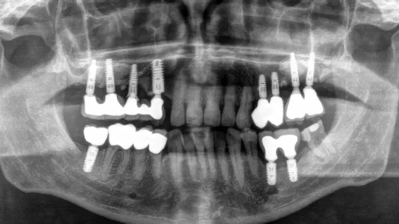
Full jaw reconstruction with NOVAMag® membrane Dr. Piero Papi, IT In this clinical case, a patient with critical state of periodontitis and extreme bone atrophy underwent full jaw reconstruction of the maxilla using cerabone® +HyA combined with NOVAMag® magnesium metal membrane. The procedure involved extensive bone augmentation to restore the jaw’s structural integrity and prepare it for implant placement. The xenograft material provided the necessary bone volume, while the magnesium membrane offered mechanical strength and supported the healing process by gradually resorbing, eliminating the need for secondary surgery. This approach ensured a stable, long-lasting foundation for dental implants. Subsequently, after 6 months five implants were placed and a fixed maxillary full arch was used to replace all the teeth in maxilla.
-
Replacement of four failing maxillary incisors due to chronic infection and external root resorption Dr. Ross Cutts, UK This lovely lady patient was referred to our clinic due to her failing upper central and lateral incisor teeth following a traumatic event many years previously. The upper centrals were splinted following mobility issues associated with the external root resorption and the upper left lateral incisor tooth had an area of chronic apical pathology giving a poor long-term prognosis. The upper right lateral incisor tooth whilst appearing healthy had some periodontal mobility also giving it a poor long-term prognosis so the patient requested to replace the upper incisor teeth with a fixed implant retained bridge solution.
-
Simultaneous lateral sinus lift and implantation of 2.6 and 2.4 with cerabone® +HyA and collprotect® membrane Dr. Alejandro Signorio The patient presented with missing teeth 2.4 and 2.6. In zone 2.6, the bone height was insufficient to place an implant of a normal size. It was decided to perform a sinus lift through a lateral window with cerabone® +HyA and collprotect® membrane for zone 2.6 and to place implants in both areas simultaneously. After osseointegration, the implants were restored with two screw-retained, cemented single crowns. After 2.5 years, the soft and hard tissues are stable and in good condition.
-
Delayed implant placement with NOVAMag® membrane in the mandible Prof. Werner Zechner A patient with a pronounced atrophic bone in the area of tooth #45 & #46, following tooth extraction several years ago, requested an implant supported solution. As the bone volume was insufficient for primary implant placement, a delayed implantation after alveolar crest regeneration was planned. Initially, the site was augmented with cerabone® in combination with autologous bone in 1:1 ratio. A NOVAMag® membrane was cut to fit the defect and shaped into the desired shape. Upon placing the bone graft, the magnesium membrane was folded over the augmented site and fixed with titanium screws both lingually and buccally. After 6 months of uneventful healing, excellent soft tissue condition were reported. Upon opening the flap for implant placement, the xenograft particles were integrated into the bone and a high quality and sufficient volume of regenerate bone was documented. The formation of new bone showed a dense and highly vascularized bone. Subsequently, 2 months after implant and check-up the patient was sent back to the referring prosthodontist for the final restoration.
-
Immediate implantation after fracture of 2.4 - soft tissue management using mucoderm® Dr. Sileno Tancredi A patient presented with a fracture of 2.4. The tooth was extracted and an implant was placed immediately in the socket together with bone grafting with cerabone® +HyA to fill the gap around the implant neck. Submerged implant placement was performed and the implant closed using a cover screw. Afterwards, mucoderm® was positioned slightly below the marginal gingiva and sutured with simple sutures to stabilize it. The matrix was intentionally
-
Immediate implant placement with Shield technique at the aesthetic area Dr. Massimo Frosecchi This clinical case represents a three-dimensional bone regeneration of an atrophic tooth in the aesthetic region. The hopeless tooth was extracted and immediate implant placement was performed. Initially, the implant was placed and the novel NOVAMag® membrane was applied to regenerate the missing buccal bone wall. The fully resorbable magnesium membrane was cut to fit the morphology of the defect and was placed between the periosteum and the bone without fixation. In order to augment the bone around the implant, cerabone® was applied and the wound was primary closed. The clinical situation after 6 months shows satisfying results – very stable bone and soft tissue conditions.
-
Reconstructive peri-implantitis treatment using photodynamic therapy and cerabone® +HyA Dr. Dragana Rakašević, Prof. Dr. Aleksa Markovic, Dr. Iva Mijailovic, Dr. Tijana Misic Prosthetic work: Prof. Dr. Miodrag Scepanovic The initial diagnosis revealed a peri-implant pocket depth (PPD) of 8 mm together with evident bleeding on probing (BOP). The radiographic and intra-operative examination confirmed a circumferential bone defect with a bone loss of approximately 2 mm. The too buccally position of the implant was considered one of the possible risk factors for the onset of peri-implantitis. PPD reduced to 3 mm six months post-surgery and one year after the treatment the clinical outcome was found stable (PPD of 3.5 mm) without sings of further bone loss or BOP. Further, keratinized mucosa width and thickness significantly increased one year after surgery. Three months postoperatively, the patient received a new screw-retained implant-supported crown. Additionally, the patient received a new full ceramic crown due to the absence of interproximal contact between premolars.
-
Full maxillary reconstruction with maxgraft® bonebuilder Dr. Christian Hilscher This case demonstrates significant bone resorption with the loss of buccal walls in the 14-24 region, along with substantial three-dimensional bone defects resulting from periimplantitis. Following the surgical removal of four implants and stabilization of the blood coagulum using collacone®, a DVT image was taken for CAD/CAM planning following a six-month healing period. For augmentation, four maxgraft® bonebuilder blocks were utilized, along with external bilateral sinus floor elevation using a combination of autologous bone and maxgraft® cortico-cancellous granules. The Schneiderian membrane was protected with several Jason® membrane, and the augmentation area was fully covered with additional Jason® membranes and PRF matrices. After seven months, successful implantation was carried out. The biopsy taken at this time point shows the remodeling of the allogenic graft (star) into patient’s new bone (arrow). 4 years radiological control shows satisfying, stable outcome.
-
Management of a Late Ti Mesh Exposure in the Aesthetic Area using Jason® membrane & cerabone® in a Sausage Technique approach Dr. Xavier Uriarte This case demonstrates the complete management of a complication in the aesthetic area, i.e. a late exposure of a Titanium Mesh. After the removal of the titanium mesh and soft tissue healing, Guided Bone Regeneration (GBR) with simultaneous implant placement was performed. GBR was carried out using the Sausage Technique (cerabone®, mandibular autograft and Jason® membrane). Prosthetics included a screwed provisional crown, along with a gingivectomy and five veneers. The patient was very happy with the result.
-
Posterior mandible GBR using permamem® and cerabone® +HyA Dr. Orlando D’Isidoro This clinical case shows a three-dimensional bone regeneration of an atrophic posterior mandible. Two implants were inserted and simultaneously bone regeneration was performed by means of a d-PTFE membrane (permamem®) and a 40:60 mix of bovine bone substitute with hyaluronate (cerabone® +HyA) and autologous bone chips. The membrane was fixed by mini-screws on the lingual and cortical bone plate. After 4 months the surgical re-entry was performed and the soft tissues were thickened in horizontal and vertical volume, with a porcine dermal matrix (mucoderm®) to achieve at least 3.5 mm of mucosa. Two stay-in abutments were connected according to the concept of “One abutment- One time”. At the same time a vestibuloplasty was carried on the tooth 3.7 with mucoderm®. The prosthetic work was delivered with screwed zirconia crowns cemented on t-base abutments.
-
Delayed implant placement with GBR and soft tissue augmentation at the aesthetic area - 2 years follow up Dr. Hassan Maghaireh & Dr. Victoria Ivancheva Patient presented with a missing upper right central incisor (UR1). This tooth was removed long time ago and there were signs of bone loss and resorption due to the bone remodelling. Patient was also undergoing orthodontic treatment due to the loss of mesio-distal space. After implant placement, GBR was performed using a 4:1 mix of maxgraft® and cerabone® shaped to create convexity and covered with Jason® membrane followed by mucoderm® to support soft tissue regeneration. Clinical situation after 2 years follow up shows satisfying stable bone and soft tissue conditions.
-
Large horizontal augmentation using the fully resorbable metal NOVAMag® membrane & cerabone® Dr. David Botond Hangyási Substantial bone resorption around tooth 27 (FDI notation system), led to root exposure and furcation involvement. Pre-operative x-ray confirmed bone resorption around the tooth. Tooth extraction resulted in a severely compromised buccal wall. NOVAMag® membrane was cut and bent to shape, and positioned to bridge the defect over the buccal wall. A convexity was created using the membrane to substantiate a large horizontal bone gain, and the defect was filled with cerabone® granules. A second piece of NOVAMag® membrane was cut with rounded edges and placed on top of the augmentation to retain the graft granules and to protect the defect space from collapse. The site was then closed with primary intention using a soft tissue graft harvested from the palate. After a five and half month healing period, reentry revealed a dense and well vascularized bone that supported the insertion of a dental implants in position 26 and 27 (FDI notation system). The original shape of the augmentation had been retained, demonstrating the capability of the NOVAMag® membrane in combination with cerabone for maintaining volume for bony regeneration.
-
Peri-implant soft tissue thickening after ridge augmentation with cerabone® and Jason® membrane Dr. Guilherme Moreira The patient presented with mobility at tooth 11, the x-ray tomography revealed a root fracture. For permanent reconstruction of the missing bone, augmentation with cerabone® and Jason® membrane was performed. collacone® was placed on top of Jason® membrane to provide additional protection since the overlying soft tissue was not completely closed. In a second step, the soft tissue was thickened with mucoderm® directly after implant placement. The follow-up shows a stable result with good emergence profile.
-
Sinus lift with additional horizontal augmentation – A 5-year follow-up Dr. Tomislav Krhen Patient presented with a failing bridge on the right distal part of the maxilla with periodontally compromised tooth 15. A two-step surgery with bone augmentation and sinus lift procedure, followed by implant placement was planned. Sinus lift procedure was performed using cerabone® mixed with autogenous bone and Jason® membrane. In addition, horizontal ridge augmentation was performed at site 15 as well as overcontouring at site 13-12. Clinical and radiological follow-up 5 years post-operative shows stable bone volume with no gum recession, minimal pocket depth on probing, no BOP.
-
Synthetic approach for Socket Preservation ****Dr. Erick Mota A fresh extraction site in the posterior maxilla was regenerated with the aim of an implant-supported restoration using fully synthetic biomaterials. In an open healing approach, permamem® was left exposed to the oral cavity for 21 days before removal. The re-entry six months post-surgery exposed a well vascularized bone tissue, which was sufficient in width and height for the placement of a dental implant. Altogether, the case demonstrates that synthetics can be a solution to achieve predicable bone regeneration in situations where an implant cannot be placed immediately.
-
GBR in aesthetic zone with maxgraft® and Jason® membrane - 5 year follow up Dr. Andoni Jones A healthy 55 year old male patient had a tooth extracted due to a failed endodontic treatment three months previously. During implant placement there was an evident buccal bone defect, and an apicoectomy of the UL1 was done simultaneously. A guided bone regeneration was done using maxgraft® cortico-cancellous granules and Jason® membrane. Three months later the implant was restored. At 5 year follow up, the radiological scan shows a satisfying, stable outcome in the aesthetic zone.
-
Maxillary sinus augmentation using cerabone® +HyA – A 2-year follow-up ****Dr. Jordi Gargallo-Albiol, Spain The patient presented with an initial residual bone height of the sinus floor of less than 2 mm. Using the lateral approach in a two-stage procedure, the missing bone volume was augmented with cerabone® +HyA, which allowed for an efficient filling of the elevated sinus, thanks to the stickiness of the material. The two-year result demonstrates a stable clinical situation, both in terms of hard and soft tissue.
-
Treatment of vertical bone loss by using cerabone®, autologous bone and Jason® membrane ****Prof. Rafael Ortega Lopes The patient (woman, around 30 years) presented with severe pain in the lateral incisor and a deficient adhesive provisional. Bruxism resulted in canine loss and premature contact in the lateral incisor. The CBCT scan indicated advanced vertical bone loss. For augmentation cerabone® was mixed with autologous bone (ratio 1:1) and covered with Jason® membrane. Due to its high stability cerabone® provides optimal support of the newly formed bone. Jason® membrane is characterized by a long barrier function and high tear resistance. The clinical follow up after 3 and 4 years demonstrates an aesthetic and stable result.
-
Reconstruction of Anterior Maxilla with maxgraft® bonebuilder – a 5 years follow-up Dr. Buket Han Patient had lost her teeth as a result of periodontitis (caused by the very often decementation of her upper bridge). CBCT data showed a horizontal and vertical bone loss in the anterior part of her upper jaw. An allogeneic customized bone block, maxgraft® bonebuilder, was the treatment of choice because of a prior failed autogenous surgery in the mandible. The customized bone block was contoured with cerabone® and covered with Jason® membrane. After 6 months, three implants were placed. 5-year follow up demonstrates a stable and satisfying outcome.
-
Posterior socket preservation using maxgraft® and permamem® – a 2-year follow up ****Dr. Cobi Landsberg & Prof. Nitzan Bichacho (Prosthetic work) A periodontally compromised, hopeless tooth in the posterior maxilla was extracted and socket preservation was performed using cortico-cancellous maxgraft® granules and permamem®, which was left exposed to the oral cavity. Even though the membrane protected the socket for only 1 week, a complication-free, complete soft tissue healing was achieved after 2 months. At the 6-month reentry the gained and maintained bone volume allowed for the placement of an implant. At the installation of the healing cap 4 months later the implant was found to be well osseointegrated. The 2-year follow up demonstrates a stable and satisfying outcome.
-
Treatment of a double root fracture in the aesthetic zone with cerabone® Dr. Jose Manuel Abarca After a fall while fishing the patient suffered a root fracture in two incisors. Careful extraction of the fractured teeth and roots allowed immediate placement of two implants. For filling the gap between implant and bone wall cerabone® granules were used. Its high volume stability makes it the ideal choice for permanent support of the ridge shape. Two-years follow-up shows a stable bony situation and a very aesthetic treatment result.
-
Maxillary bone reconstruction in the anterior area with cerabone® and Jason® membrane Pierre-Yves Gegout and Prof. Olivier Huck Patient presented with loss of interdental papilla and gingival inflammation in the aesthetic zone. After extraction of teeth 12 and 11, the area was rinsed with chlorhexidine and saline. The distinct ridge defect was then reconstructed immediately using cerabone® and Jason® membrane. Thanks to the long-term volume stability of cerabone®, the clinical situation presents itself stable with a fixed bridge after 22 months follow up.
-
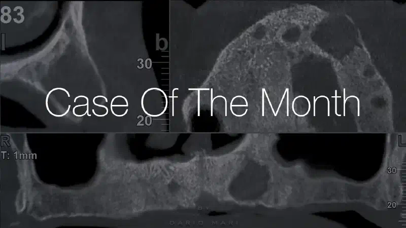
Treatment of a maxillary cyst with cerabone® and Jason® membrane Dr. Dario Mari A female adult patient was complaining about swelling and pain in the upper left maxilla. She could not even wear her denture. CBCT confirmed a huge cyst in the left cuspid area, but other minor cysts were present in other sites of the maxilla. After full thickness flap from molar to molar, the lesions were excised with a sharp surgical spoon to keep them as intact as possible. The residual bone cavities were curetted and rinsed with saline water and antibiotics (Metronidazole). GBR procedure was performed filling the gaps with cerabone® (granule size 1-2mm), covered by a Jason® membrane, which was fixed by titanium pins. The flap was sutured with PTFE sutures. The histology confirmed the initial diagnosis of residual cysts after tooth extraction. After 9 months healing time, four implants were placed.
-
Sinus lift and lateral bone augmentation with cerabone® and maxgraft® Dr. David Chávarri Prado Due to periodontal disease, the patient (65 years) had a failure of the fixed prosthesis in the second quadrant and presented with severe bone atrophy, both in height and width. A lateral sinus lift and simultaneous GBR were performed using a 50/50 mix of cerabone® and maxgraft® granules and PRGF. The augmented area was covered and stabilized with Jason® membrane, which has a naturally long barrier function and high tear resistance. Six months later, at the re-entry very good bone reconstruction was achieved.
-
Horizontal ridge augmentation with maxgraft® cortico, cerabone® and collprotect® membrane Eleni Kapogianni A missing dentition in the mandible was reconstructed with maxgraft® cortico and cancellous allogenic granules. The site was covered with a collprotect® membrane and was re-entered after 5 months. Two implants were placed in area 44 and 46 and then covered with cerabone® for optimal volume stability and aesthetic outcome (Relining technique).
-
Block augmentation in the aesthetic zone with maxgraft® and cerabone® Dr. Frank Kloss After fracture of the left maxillary incisor and loss of buccal wall an extensive bone defect was reconstructed by onlay block grafting with a cancellous maxgraft® block and relining with cerabone® granules. Six months after augmentation successful implantation was performed and again covered with cerabone® for long-term stability of the gained horizontal width.
-
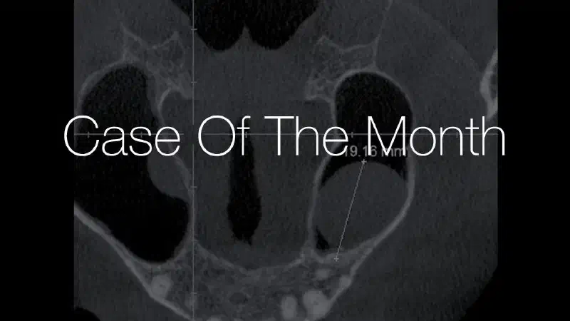
Maxillary sinus cyst removal using the Crocodile Technique and subsequent lateral sinus lift with cerabone®, maxgraft® and Jason® membrane Dr. Cristian Scognamiglio, Dr. Alessandro Perucchi After extraction of a severely compromised tooth 25, a maxillary sinus cyst with a size of 19 mm had to be removed before implantation and sinus floor elevation. For removal, a lateral sinus window was prepared, and the cystic content was aspirated through the membrane. The cyst was then retrieved through a cut in the Schneiderian membrane according to the Crocodile Technique. After suturing the perforation, the sinus membrane was protected with Jason® membrane which is a native pericardium membrane with long barrier function. Eight months later, sinus lift and implantation were successfully performed.
-
Excellent aesthetic result of buccal augmentation with mucoderm® and maxgraft® after immediate implant placement - 3-years follow-up Dr. Algirdas Puišys After atraumatic tooth extraction the implant was immediately placed and the gap between buccal bone wall and implant was filled with cancellous maxgraft® granules. For soft tissue augmentation the collagen matrix mucoderm® was used: A subperiosteal buccal tunnel was prepared at the extraction site and mucoderm® was inserted and fixed by suturing. An excellent aesthetic outcome was achieved as also shown by the results in a clinical study published in 2022. https://pubmed.ncbi.nlm.nih.gov/35324053/
-
cerabone® and mucoderm® for immediate implantation in aesthetic area Dr. Daniel Robles For an optimal aesthetic result, cerabone® and mucoderm® were used for hard and soft tissue augmentation during immediate implant placement in the front tooth region. The high volume stability of cerabone® ensures permanent support of the bone, while mucoderm® is a perfect collagen matrix for soft tissue thickening.
-
Socket Preservation with maxresorb® and mucoderm® Dr. Massimo Frosecchi maxresorb® is a synthetic bone substitute with excellent handling characteristics. Due to its biphasic composition, it promotes bone formation while supporting stability of the augmented area. In a lot of indications it is a valid alternative to xenografts, but avoids ethical or religious conflicts. In the shown case maxresorb® was used in combination with the soft tissue graft mucoderm® for socket preservation. The case shows good bone and soft tissue healing.
-
Horizontal ridge augmentation with permamem®, cerabone® and autologous bone chips Dr. Stavros Pelekanos Great bone regeneration was achieved after horizontal ridge augmentation using permamem®, cerabone® and autologous bone chips. permamem® is a synthetic membrane with superior space maintaining properties compared to collagen membranes. Due to its dense structure and smooth surface, soft tissue attaches only superficially which makes it easy to remove the membrane after healing.
-
GBR with simultaneous implant placement and widening of KM with mucoderm® Dr. Marko Blašković Eight weeks after tooth extraction GBR was performed with simultaneous implant placement. The combination of cerabone® with autologous bone and Jason® membrane led to a beautiful restoration of the missing bone volume. Additionally, broadening of the keratinized mucosa was required but the patient refused to undergo a FGG grafting procedure. Thanks to mucoderm® (acellular dermal collagen matrix) the treatment was carried out successfully and a greater width of keratinized mucosa was achieved.
-
Ridge augmentation in the mandible with maxgraft® bonebuilder Dr. Reto Morger A 44-year-old female patient presented for implant-prosthetic treatment in the third and fourth quadrant. Since the area had not been restored for more than 20 years, an extensive horizontal and vertical augmentation was required prior to implant placement. The mandible bone was reconstructed using two maxgraft® bonebuilder blocks. The customized allogenic bone blocks fitted perfectly and were covered with a Jason® membrane. After 6 months healing time implants were installed at position 35/36 and 46. 1 year follow up shows good stable clinical outcome.
-
Immediate implant placement in the maxilla with contour GBR Dr. Konstantinos Loukas In this clinical case a combination of cerabone® and autologous bone chips was used to fill the gap between implant and socket wall and to contour the ridge. The high volume stability of cerabone® makes it the ideal choice in the aesthetic area.
-
Simultaneous implantation and ridge augmentation using maxgraft® cortico, maxgraft® granules and mucoderm® Dr. Algirdas Puišys This case shows 5 years outcome after simultaneous implantation and ridge augmentation with allogenic bone plate maxgraft® cortico. Stable space was created filled with maxgraft® granules then covered with mucoderm® to maintain an aesthetic outcome in the long term.
-
GBR in the aesthetic zone with cerabone® and Jason® membrane Dr. Ross Cutts In the anterior region bony support of the soft tissue is essential to achieve optimal aesthetic results. In this case cerabone® was used for augmentation, the material shows ultimate volume stability and thereby supports a predictable long-term clinical outcome. The augemented area was covered with Jason® membrane which is especially advantageous for the regeneration of larger defects due to its naturally long barrier function.
-
Immediate implant placement using maxresorb® Dr. Alejandro Signorio In a lot of indications the synthetic bone substitute material maxresorb® is a good alternative to materials of animal origin. Here it was used to fill the gap around an immediately placed implant. The case was followed up to 4 years and shows a good clinical result with stable implant integration.
-
Reconstruction of maxillary ridge with maxgraft® block Amit Patel A severe maxillary atrophy was augmented using a maxgraft® block and bovine bone granules. maxgraft® is a sterile allograft product with preserved biomechanical properties. The block was cut into slices and fixed with osteosynthesis screws. For filling up the voids bovine bone granules were used and the augmented area was covered with a collagen membrane. Due to the natural human bone structure and the preserved collagen in maxgraft® blocks a fantastic horizontal bone regeneration was obtained.
-
Reconstruction of a single tooth defect with the shell technique ****Dr. Andoni Jones: A considerable horizontal defect was grafted with maxgraft® cortico, maxgraft® cortico-cancellous granules and Jason® membrane in a single tooth gap. The maxgraft® granules were put in the gap and over the strut buccally restoring the missing buccal plate. The combination of biomaterials achieved an increase in horizontal dimension and perfect volume maintenance of the graft (1 year follow up).
-
Vertical bone augmentation and broadening of attached gingiva using cerabone®, permamem® and mucoderm® ****Dr. Ruggero Naimoli: Starting from a challenging initial situation with pronounced vertical and horizontal bone defect a good bone regeneration was achieved by using cerabone® granules, permamem® and Jason® membrane. After placing two implants, augmentation with the soft tissue graft mucoderm® allowed broadening of the attached gingiva.
-
Bone regeneration and implant-supported rehabilitation of a periodontally involved incisor Dr. Andres Eslava Vanegas: A mandibular front tooth with an endo-perio lesion was extracted and immediate implant placement in conjunction with GBR was performed. cerabone® in combination with autologous bone chips and PRF was used, which was covered by mucoderm®. The 3-year outcome demonstrates maintenance of the bone volume and a stable soft tissue situation.
-
Hard and soft tissue management in immediate restoration in the aesthetic area with partial edentulism Dr. Massimo Frosecchi: The clinical case shows the removal of both central incisors due to a fistula and abscesses. As the incisors were supporting a bridge, the bridge between lateral incisors and canines were cut and immediate implant placement for a new 4-unit bridge was planned. A high esthetic outcome was achieved at the final end.
-
Shell Technique with augmentative relining with cerabone® and Jason® membrane (Split Mouth with maxgraft® cortico and autologous bone plates) Dr. Jochen Tunkel: This case is part of a Split-Mouth-Study, patient had a bilateral bone defect requiring three-dimensional augmentation and was treated with autogenous and allogeneic bone plates (maxgraft® cortico) in 4th and 3rd quadrant respectively. Augmentative relining with a xenogeneic bone substitute material (cerabone®) and collagen membrane (Jason® membrane) minimized resorption processes and maintained bone volume in the long term.
-
mucoderm® for regeneration following removal of an odontogenic fibroma Prof. Dr. Dr. Daniel Rothamel The clinical case shows an odontogenic fibroma that was growing for years in a 57-year-old man (a dental phobics). mucoderm® has been successfully applied following resection of the fibroma to cover the open wound and to fully regenerate the soft tissue including the keratinized mucosa.
-
Ridge augmentation with maxgraft® bonebuilder and vestibuloplasty with mucoderm® in the maxilla Dr. Oliver Blume: A patient presented with a complex maxillary defect in area 14-16. The combination of maxgraft® bonebuilder and mucoderm® for vestibuloplasty during implant placement enabled the regeneration of vital bone tissue and good aesthetic appearance without the need of harvesting autologous hard and soft tissue.
-
Regeneration of a 9 mm vertical bone defect with cerabone®, autologous bone and S-PRF Dr. Andrés Eslava Vanegas: A patient presented with suppuration and pronounced bone loss around 2 implants placed 5 years ago in another dental clinic. Two months after implant extraction, the extent of the bone defect was 9 mm vertically. After augmentation with cerabone® (mixed with autologous bone and S-PRF) an immense bone gain was achieved and allowed for stable implantation of two new implants.
-
Immediate implant placement and periimplant bone augmentation using cerabone® Dr. Ricardo Kern: Tooth fracture due to trauma in the anterior maxilla was rehabilitated by immediate implant placement and periimplant bone augmentation using cerabone®. The approach optimally supports periimplant hard- and soft tissue regeneration after tooth removal as demonstrated by a stable radiographic bone level and a sound stability of the marginal soft tissues.
-
Smile rehabilitation in the anterior maxilla with maxgraft® bonebuilder Dres. A. Dagba and J. Mourlaas: A patient presented with an unharmonious smile due to missing teeth in the upper right maxilla. After bone augmentation with maxgraft® bonebuilder the patient received a fixed prosthetic solution on three implants. The final radiograph with a two year follow up revealed a good bone stability and favorable adaptation of the prosthesis, giving the patient a successful smile rehabilitation.
-
Full arch GBR using cerabone®, maxgraft® and Jason® membrane with simultaneous implantation of 6 Straumann® implants Dres. A Signorio and G. Molina: The outstanding restoration of the buccal contour using a combination of the biological potential of maxgraft® with the volume stability of cerabone®, allowed the complete rehabilitation of the patient who presented a highly atrophied fully edentulous maxilla with implant supported bridges with screw retention instead of a full arch screw retained prosthesis.
-
GBR of the edentulous maxillary ridge using permamem®, cerabone® and autologous bone chips Dr. Viktor Kalenchuk: The „case of the month“ is a new category in the indication matrix, which highlights every month a clinical case, which distinguished itself by the clinical results or the treatment concept in combination with the applied botiss biomaterials. The selection of the case is based on content relevance and quality of the documentation. Apply now and share your case of the month -> product-management@botiss.com
-
Full bone regeneration in extraction socket augmented with maxgraft® and Jason® membrane Dr. Cobi Landsberg: Associated with a root fracture, a patient presented with a pocket depth of 9 mm and advanced loss of palatal bone. Due to the biologic potential of maxgraft® granules in combination with the favorable properties of Jason® membrane, full bone regeneration was obtained.
-
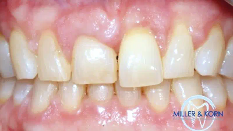
Immediate implant placement and correction of horizontal and vertical bone loss using an allograft bone ring, cerabone® and Jason® membrane Drs. Miller and Korn: A patient presented with pathologic tooth mobility and a recession associated with the upper left central incisor. Due to the application of the bone ring combined with cerabone® and Jason® membrane multiple surgeries could be avoided. The final radiograph (8 months postoperatively) revealed both horizontal and vertical augmentation of the osseous defects. Root coverage of the recession on the labial aspect of the lateral incisor was noted as well.
-
Treatment of a combined horizontal and vertical bone defect in the maxilla with maxgraft® cortico in the allogenic shell technique **Dr. med. dent.**Robert Würdinger: A severe horizontal and vertical bone defect was treated with maxgraft® cortico without the need of bone block harvesting. Additionally, an external sinuslift with cerabone® was previously performed at the defect site. Due to the excellent results with the shell-technique, no augmentative relining with xenogeneic material was needed at the end!
-
Application of mucoderm® in soft tissue management around single implants at second stage surgery Dr. Carlo De Annuntiis: The „case of the month“ is a new category in the indication matrix, which highlights every month a clinical case, which distinguished itself by the clinical results or the treatment concept in combination with the applied botiss biomaterials. The selection of the case is based on content relevance and quality of the documentation. Apply now and share your case of the month -> product-management@botiss.com
-
Entire papilla preservation technique (EPP) for the regenerative treatment of a severely compromised central incisor Dr. Serhat Aslan: The „case of the month“ is a new category in the indication matrix, which highlights every month a clinical case, which distinguished itself by the clinical results or the treatment concept in combination with the applied botiss biomaterials. The selection of the case is based on content relevance and quality of the documentation. Apply now and share your case of the month -> product-management@botiss.com
-
Horizontal/vertical GBR using cerabone®/maxgraft®, Jason® membrane/collprotect® membrane and Ti-mesh Dr. Andrew Aziz: The „case of the month“ is a new category in the indication matrix, which highlights every month a clinical case, which distinguished itself by the clinical results or the treatment concept in combination with the applied botiss biomaterials. The selection of the case is based on content relevance and quality of the documentation. Apply now and share your case of the month -> product-management@botiss.com
-
In this clinical case, a patient presented a cracked dental root in the incisor region, specifically on tooth 11, rendering prosthetic rehabilitation of the natural tooth highly challenging. In addition, the patient exhibited gingival recession at the affected site. The tomographic images confirmed the presence of an oblicual root fracture. Based on the clinical and radiographic findings, the treatment plan consisted of an atraumatic tooth extraction followed by an immediate implant placement in order to preserve hard and soft tissue architecture and optimize the aesthetic outcome.
-
Socket preservation with a short re-entry time using maxgraft®, cerabone® and mucoderm® Dr. Bartosz Matejkowski Following atraumatic tooth extraction, socket preservation was performed using a mixture of maxgraft**®** / cerabone**®** with a ratio of about 65%/35%. botiss grafter was used to rehydrate the bone graft mixture properly and apply it elegantly in the socket. The graft was covered with mucoderm**®** for open healing. The re-entry was done after only 2 months of healing. At re-entry, vital and highly vascularized bone-bed was noticed. A biopsy was harvested using a trephine for histological analysis. A bone-level implant was successfully placed and restored. Radiographs taken 7 months after implant placement show good integration of the implant and sufficient bone levels.
-
A 48-year-old female patient presented with persistent pain in the region of tooth 36. The symptoms occurred despite ongoing endodontic treatment. Clinically, there was pronounced tenderness to percussion, but no swelling or fistula formation. Cone beam computed tomography (CBCT) revealed an extensive periapical osteolysis at the distal root, along with a fine hairline crack in the same root. After thorough patient consultation regarding treatment options, the tooth was carefully extracted. Simultaneously, socket and ridge preservation were performed using cancellous maxgraft® +HyA – an allogeneic bone substitute material combined with hyaluronic acid. No membrane was used to cover the bone graft, only mattress suture. Postoperative healing was uneventful.
-
Simultaneous buccal and palatal alveolar ridge reconstruction using biodegradable magnesium membrane shield technique Dr. Akiva Elad The patient was a 65-year-old female in good general health condition. She presented with tooth 24, root canal treatment, post, core build up, and an old porcelain-fused-to-metal (PFM) crown. A vertical root fracture was present with an associated severe bone loss, including loss of both buccal and palatal plates. After the non-traumatic extraction of tooth 24, the magnesium membrane shield technique was performed (Elad et al. 2023) using NOVAMag® membrane, aiming to rebuild both buccal and palatal plates. Due to the mechanical stability of the membrane, it can be easily inserted and put in position. The defect was filled with maxgraft® (allogenic bone) and Jason® membrane (pericardium collagen membrane) placed over the top of the augmentation and the flaps were sutured.
-
Shield Technique with NOVAMag® membrane A clinical case by Dr. Massimo Frosecchi demonstrating the Shield technique using the fully resorbable NOVAMag® membrane. By preserving the natural bone structure, this technique minimizes the need for complex procedures and additional grafting, supporting successful and predictable outcomes with less surgical intervention. NOVAMag® membrane is made from pure magnesium metal and is completely resorbable. It is also characterized by exceptional strength during the critical healing phase and can withstand the pressure of the soft tissue, providing space for the bone graft.
-
Shield Technique in a case with immediate implant placement Dr. Luka Markovic Following tooth extraction in the mandibular, the clinical situation presented a severely compromised buccal wall with complete loss of the buccal plate. An implant was placed immediately into the extraction socket, and the defect was grafted with cerabone ® +HyA, whose high-volume stability and excellent biocompatibility provide a predictable and long-lasting foundation for implants. To ensure space maintenance and create an optimal environment for bone regeneration, NOVAMag® SHIELD was carefully tucked between the bone and soft tissue and secured with sutures; its strong mechanical properties and gradual magnesium resorption allow for stability during healing without the need for removal. Finally, mucoderm® was applied over the augmented site to increase soft tissue thickness. This xenogeneic collagen matrix serves as an effective alternative to autologous connective tissue grafts. Primary wound closure was achieved, ensuring stable healing and optimal conditions for regeneration. At six months, the follow-up demonstrates strong regenerative results and stable new bone formation.
-
Dr.RaphaëlBettach This case involved a complex upper jaw reconstruction in a patient presenting with multiple missing teeth and severely compromised bone volume. During the surgical phase, a mucoperiosteal flap was elevated to expose the underlying bone, revealing significant buccal bone defects across several implant sites. After careful assessment, implants were strategically placed, including in regions with vestibular bone deficiencies that required regenerative support. The defects were filled with a bone grafting material to promote new bone formation and restore volume. A folded NOVAMag® membrane was then applied to maintain space and provide structural stability. In certain areas, a collagen membrane was combined with NOVAMag® membrane to support soft tissue integration and optimize healing.
-
Dr.****Tin Crnić**** An 80-year-old patient presented with pain and mobility in tooth 25. Clinical and radiographic evaluation revealed advanced periodontal destruction and a hopeless prognosis for tooth 25, while adjacent teeth 24 and 26 tested vital and stable. Tooth 25 was extracted under local anaesthesia, revealing a buccal wall defect. To preserve the alveolar ridge, NOVAMag® SHIELD was placed and collacone® was inserted into the socket to support clot stabilization and healing.
-
Resorbable magnesium metal membrane for a sinus lift procedure - application of the NOVAMag® membrane Dr. Akiva Elad The patient, a 61-year-old male in good overall health, presented with two dental implants at positions 14 and 16, supporting a fixed dental bridge. Examination revealed movement in both vertical and horizontal dimensions, whereas peri-implantitis was indicated, with vertical bone loss up to half the height of implant 14, and implant 16 located in the right maxillary sinus surrounded by a large polyp. The dental implants were extracted atraumatically, preserving the Schneiderian membrane. A direct sinus lift with a buccal window was performed, draining and removing the sinus polyp with an aspiration needle. A magnesium membrane was placed on the alveolar bone of the buccal window, with maxgraft® and cerabone® granules applied into the buccal window to seal it off from the alveolar bone. Additional graft material was placed in the extraction site and covered with a long piece of magnesium membrane along the alveolar ridge for adequate coverage and separation from the soft tissue. A dental implant was then placed at position 14. Five months post-surgery, the bone displayed optimal hardness and structure. CBCT imaging showed 16–21 mm of newly formed bone, varying by location. Two additional dental implants were subsequently placed at positions 15 and 17.
-
Resorbable magnesium membranes and fixation screws for GBR in delayed implant placement Dr. Marko Blašković Cone beam computed tomography (CBCT) of tooth 12 (FDI notation system) revealed bone deficiency in both horizontal and vertical directions. A complete mucoperiosteal flap was raised and the site intended for augmentation was exposed. cerabone® and a small amount of locally harvested autogenous bone were mixed together and used to augment the defect. NOVAMag® membrane was used to separate the defect site from the overlying soft tissue. The membrane was secured to prevent slippage using resorbable NOVAMag® fixation screws. To achieve an optimal soft tissue profile, mucoderm® was also placed over the membrane. The clinical outcome after 3 months was satisfactory, with the labial and oral mucosas being close together, and soft tissue seen to be regenerated. After a total of 5 months of healing, CBCTs show bony healing in vertical and vestibulo-ral dimensions.
-
Rehabilitation with cerabone® and Jason® membrane after implant failure Dr. Lucas Werutsky The patient presented after unsuccessful implant treatment in region 11. Panoramic and periapical radiographs and a CBCT were taken to evaluate the bone dimensions. During surgery, fibrous tissue was removed after which the defect presented larger than expected from the tomographic examination. The implant was installed in a suitable position without fenestration. To correct the bone defect and increase the buccal bone volume cerabone® and Jason® membrane were used. cerabone® is particularly suitable to provide permanent support in the aesthetic region due to its long-term stability. The follow-up shows a successful result with stable bone reconstruction.
-
Regenerative management of an advanced endo-periodontal lesion using cerabone® +HyA and Jason® membrane Dr. Rodrigo González Terrats Description: A 50-year-old patient presented with advanced localized periodontal destruction at teeth 31 and 33, characterized by deep probing depths (10–12 mm), bleeding, suppuration, and increased tooth mobility. Tooth 31 was diagnosed with pulp necrosis and symptomatic apical periodontitis associated with a combined endo-periodontal lesion. Comprehensive initial therapy included occlusal adjustment, splinting, meticulous subgingival debridement, and root canal treatment of tooth 31. At 6-week re-evaluation, despite improved oral hygiene, residual deep defects persisted, confirming the indication for regenerative intervention.
-
This case presents a severe periodontal lesion with a 13 mm probing depth, suppuration on probing, and a deep infra-osseous defect. Clinical examination revealed that the lingual bony wall was missing, complicating regenerative management. A unilateral lingual flap was elevated with papilla preservation to ensure minimal trauma to the interdental tissues. Inflammatory tissue was carefully removed, creating a clean environment for regeneration. To reconstruct the missing lingual wall, mucoderm® was applied as a soft tissue scaffold to replace the deficient bony support. The infra-osseous defect was then filled with cerabone® +HyA , chosen for its volume stability, supporting predictable bone regeneration. Primary closure was achieved using horizontal mattress and single interrupted sutures. Post-operative imaging included immediate post-op radiograph for baseline evaluation and 6-month periapical radiograph, confirming stable bone augmentation. Clinically, probing depths reduced from 13 mm initially to 2 mm at 6 months, with no soft tissue recession, demonstrating successful regeneration and soft tissue management.
-
Regeneration of a compromised extraction socket with NOVAMag® SHIELD Dr. Róbert Kemper Description: A 67-year-old female patient with no significant medical history presented with moderate pain associated with the upper second premolar. Clinical examination revealed a vertical root fracture visible from the buccal aspect. The preoperative radiograph revealed a pronounced buccal bone deficiency, which was then confirmed post-extraction with completely missing buccal bone. A regenerative approach using NOVAMag® SHIELD was selected. Following atraumatic tooth extraction, the alveolar socket was carefully debrided. Subsequently, the gingival tissues were detached from the underlying bone using a tunneling knife to create a pouch for NOVAMag® SHIELD insertion. The SHIELD was adapted to match the morphology of the defect and inserted in a flapless matter. The implant was subsequently placed, and the jumping gap was grafted using a xenograft material. Immediate loading was then performed with a temporary screw-retained crown. The early postoperative healing phase was uneventful, with stable soft tissue healing and no reported complications during the immediate clinical follow-up period.
-
Reconstruction of the alveolar buccal wall in a compromised extraction socket using NOVAMag® SHIELD and cerabone® Dr. Alfonso Caiazzo A patient presented with a fractured maxillary first molar (tooth 16) requiring extraction. Due to significant bone loss, a staged approach was planned, performing bone augmentation followed by delayed implant placement. Following tooth extraction, NOVAMag® SHIELD was placed on the buccal side in a flapless approach to replace the missing wall and provide space for proper bone regeneration in the socket. The extraction socket was grafted with cerabone® followed by folding the external apical part of NOVAMag® SHIELD over the alveolus to provide structural reinforcement. The soft tissue was sutured, intentionally leaving the folded portion of the shield exposed.
-
Predictable socket preservation with collacone® and NOVAMag® SHIELD Prof. Dr. Sigmar Schnutenhaus A patient presented with a fractured, root canal-treated tooth 24 that was deemed non-restorable. To preserve the ridge architecture for future implant placement, a socket preservation procedure was planned. The preoperative radiograph revealed a pronounced buccal bone deficiency. Following atraumatic extraction and curettage of the socket, a collacone**®** cone was inserted into the alveolus. NOVAMag® SHIELD was then trimmed and adapted to the defect after preparation of a subperiosteal buccal pouch. The magnesium membrane was positioned to stabilize the buccal contour and maintain the regenerative space. Stabilization sutures were placed and an open healing protocol was performed to support the development of optimal keratinized soft tissue.
-
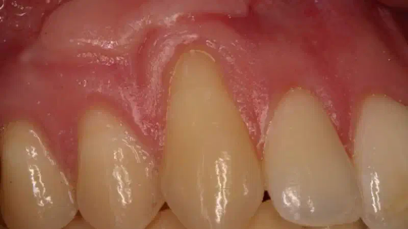
Peri-implant Soft Tissue Phenotype Modification Using Xenogeneic Collagen Matrix Dr. Ahmed ElBana A 40-year-old healthy male presented with a peri-implant soft tissue defect characterized by a thin soft tissue phenotype and a recessed mucosal margin. Soft tissue augmentation was indicated to enhance peri-implant tissue stability and esthetics. A provisional crown on a custom titanium abutment was fabricated with an emergence profile replicating the recessed gingival margin. This allowed controlled soft tissue conditioning and facilitated subsequent coronal repositioning of the peri-implant mucosa.
-
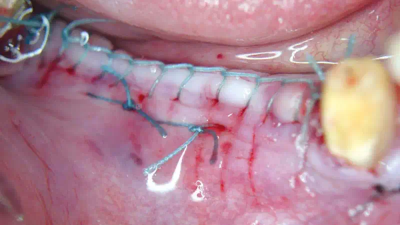
In this clinical case, a patient presented with an edentulous posterior mandible in the region of teeth 45 and 46, characterized by reduced soft tissue volume, a flattened ridge contour, and a thin mucosal phenotype. Following flap elevation, sufficient vital bone was identified, allowing implant placement without the need for hard tissue grafting. Nevertheless, reduced soft tissue thickness was noted, substantiating the indication for soft tissue augmentation. Implant placement was performed using a digitally guided, prosthetically driven approach. Due to the evident soft tissue deficiency, a native collagen matrix ( mucoderm® ) was applied on the buccal aspect to increase tissue thickness, enhance contour, and support long-term peri-implant stability. Wound closure was achieved using absorbable sutures.
-
NOVAMag® Shield technique & immediate implant placement in anterior maxilla Dr. Erick Mota A 28-year-old patient came to the consultation with a fracture of tooth 21. The previous root canal treatment and resin restoration were unsatisfactory. Due to the fracture and injury, the patient had no vestibular bony plate. Implant placement was performed with a conical connection using guided surgery. An intermediate abutment (One abutment one time) was placed to distance the prosthetic connection from the implant. To restore aesthetics and maintain tissue support, a provisional PMMA crown was placed, and the alveolar ridge was reconstructed using NOVAMag® membrane, connective tissue graft, and maxgraft®.
-
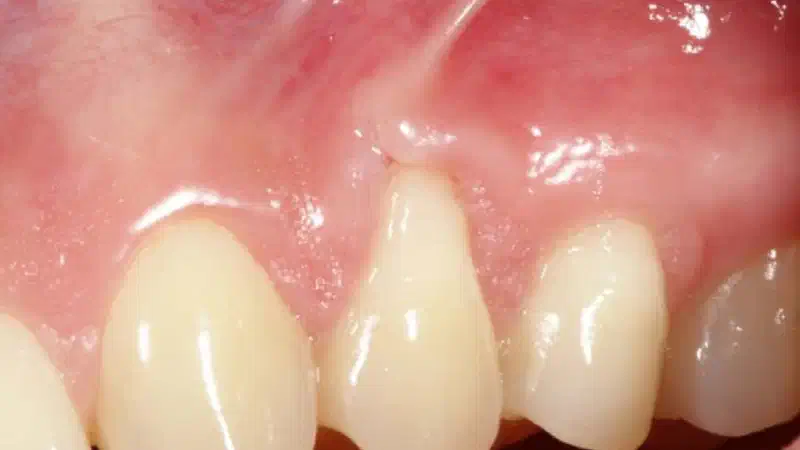
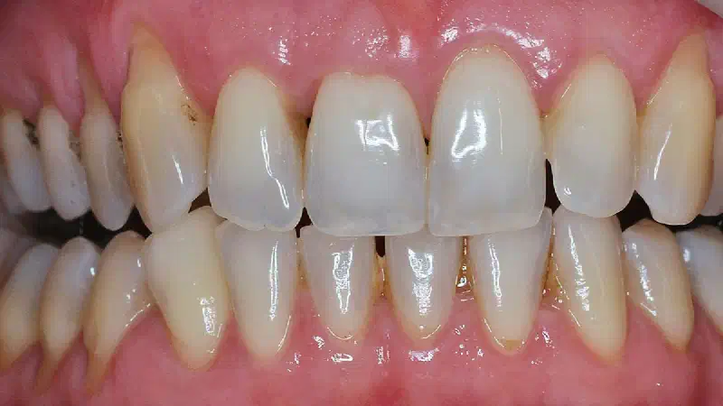
Multiple recession coverage with mucoderm® and Straumann® Emdogain® - 4 year follow-up Prof. Giovanni Zucchelli & Dr. Dr. Martina Stefanini Initial SituationThe patient presented with multiple adjacent gingival recession defects requiring predictable root coverage and soft tissue augmentation. These defects posed a challenge due to the extent and number of recessions, increasing the clinical difficulty of achieving long-term coverage and stable soft tissue volume. TreatmentA coronally advanced flap procedure was performed in combination with mucoderm® , an acellular collagen matrix, and Straumann® Emdogain®, an enamel matrix derivative gel, applied to the exposed root surfaces before placement of the matrix. The approach aimed to enhance revascularization, support soft tissue regeneration, and increase gingival thickness without harvesting autologous connective tissue.
-
Mucosal thickening around bone level implants Dr. Algirdas Puišys Initial SituationA patient presented with a missing posterior tooth and a thin peri-implant soft tissue biotype, characterized by limited vertical mucosal thickness. Thin, soft tissue is associated with an increased risk of crestal bone loss and compromised long-term implant stability. TreatmentA bone-level implant was placed, and simultaneous soft-tissue thickening was performed using the collagen matrix mucoderm® to enhance peri-implant mucosal volume. The matrix was positioned to increase tissue thickness and support stable soft-tissue healing around the implant.
-
mucoderm® for full arch reconstruction of insufficient vestibular depth and lack of keratinized tissues Dr. Balint Molnar & Prof. Peter Windisch Initial Situation The patient presented with a severely reduced vestibular depth and inadequate keratinized mucosa in the edentulous maxilla, complicating prosthetic rehabilitation and long-term peri-implant health. The lack of attached gingiva and vestibular height posed challenges for stable soft tissue support and adequate prosthesis seating. TreatmentA full-arch reconstruction was performed using multiple implants combined with soft-tissue augmentation using mucoderm®, an acellular collagen matrix, to increase keratinized tissue width and vestibular depth. The matrix was placed under a mucoperiosteal flap and secured to promote revascularization and stable integration without the need for autologous tissue harvesting.
-
Management of Periimplantitis and Implant Replacement with Guided Bone Regeneration with NOVAMag® membrane and mucoderm® Dr. Marko Blaskovic A patient presented with periimplantitis affecting an implant placed four years ago. The CBCT scan confirmed the presence of periimplant bone loss around the implant. The initial treatment plan involved the removal of the crown, followed by disinfecting the implant surface while simultaneously regenerating the periimplant bone. However, due to an unexpected implant fracture, the surgical plan had to be altered, necessitating the extraction of the affected implant. The site was thoroughly cleaned after implant removal, and the wound was closed to promote healing. Subsequently, a new implant was placed in the same area. A double layer of NOVAMag® membrane was positioned at the buccal site and fixated with NOVAMag® fixation screws. Bone augmentation was carried out using cerabone® mixed with autologous bone to ensure proper regeneration. To support soft tissue healing, mucoderm® was placed over the augmented site, followed by primary wound closure. After 6 months, implant uncovering took place.
-
Management of buccal deficiency with NOVAMag® SHIELD for enhanced implant stability Dr. Alfonso Caiazzo In this case, the initial socket preservation was performed using suboptimal biomaterials, resulting in insufficient bone regeneration. A preoperative CBCT assessment was conducted to evaluate the socket condition and surrounding bone structure. At the six-month re-entry, insufficient regeneration of the buccal plate was observed, compromising the available bone volume for implant placement. To address this deficiency, NOVAMag® SHIELD was introduced to reconstruct the buccal plate. Thus, an implant was placed followed by a membrane placement between the periosteum and the remaining buccal bone without fixation. Additional bone augmentation was performed to facilitate guided bone regeneration.
-
Long-term outcomes of horizontal ridge augmentation with GBR using cerabone® - A 6.5-year follow-up case Dr. Ketkee P Asnani A 45-year-old healthy female presented with a dislodged bridge in the lower right posterior mandible. Clinical examination revealed compromised abutment health, making preservation of the existing prosthetic solution unfavorable. Considering the patient’s age, excellent systemic health, and desire for a long-term fixed rehabilitation, a treatment plan involving horizontal ridge reconstruction followed by implant placement was established. Pre-operative CBCT analysis demonstrated significant horizontal bone loss in the affected region. The treatment objective was to restore ridge width using a combination graft composed of autogenous bone and cerabone**®** .
-
Lateral sinus floor augmentation with cerabone® with 4-year follow-up Dr. Vladimir Garcia Lozada The patient presented with an initial residual bone height of the sinus floor of 5.10 mm. Using the lateral approach in a one-stage procedure, the bone volume dimension was augmented with cerabone® which allowed for an efficient filling of the elevated maxillary sinus in conjunction with implant placement. The four-years result demonstrates a stable clinical situation, in terms of augmented hard tissue and implant efficient stability.
-
Lateral block augmentation in the posterior maxilla with cancellous maxgraft® block https://pubmed.ncbi.nlm.nih.gov/38392246/ Dr. Alexandre Perez, Prof. Tommaso Lombard INITIAL SITUATION A 53-year-old male presented with a severe horizontal bone loss in positions 13 and 14 that was associated with the removal of an included canine, performed 3 months prior. Diagnostic CBCT indicated a residual vestibular-palatal alveolar thickness of 2 mm in the edentulous premolar area. TREATMENT A two-staged implant therapy was performed comprising a lateral bone block augmentation using maxgraft® cancellous block covered with a collagen membrane followed by implant placement in positions 13 and 14 after 5 months of augmentation.
-
Immediate implantation with the Shield technique in the aesthetic zone Dr. Akiva Elad A 62-year-old male patient in good overall health presented with a horizontal oblique root fracture in tooth 21, accompanied by severe bone loss, including the complete loss of the buccal cortical plate, while the palatal cortical plate remained intact. A non-traumatic extraction of tooth 21 was performed, followed by bone augmentation using the Shield technique to reconstruct the buccal wall. Given the aesthetic zone location, the implant was immediately restored with a provisional crown. Four months postoperatively, the bone defect showed excellent regeneration, including a fully restored buccal cortical plate. The implant remained stable, with well-healed soft tissues, indicating a successful recovery.
-
Immediate implantation in the posterior mandible Block_Veras_1.jpg Initial situation Block_Veras_2.jpg CBCT planning Block_Veras_3.jpg CBCT planning for region #46 Block_Veras_4.jpg Situation before extraction of the remaining tooth root Block_Veras_5.jpg Situation before tooth extraction Block_Veras_6.jpg Identification and marking of the future implant position Block_Veras_7.jpg After tooth root extraction Block_Veras_8.jpg Preparation of the implant bed
-
A 30-year-old patient presented with a compromised upper right central incisor (tooth 1.1) due to a root fracture. Following clinical and radiographic assessment, the tooth was deemed unrestorable. An atraumatic extraction was performed, successfully preserving the buccal bone. Immediate implant placement was with excellent primary stability. To optimize the peri-implant soft tissue contour and volume, a connective tissue graft was harvested from the palatal mucosa and placed on the buccal aspect. The small buccal gap was filled with cerabone® +HyA mixed with autogenous bone, providing excellent volume stability and enhanced handling properties due to the pronounced liquid-binding capacity of hyaluronate, which forms a sticky and malleable material for easy and precise application. This combination ensured optimal adaptation to the defect morphology and promoted effective bone regeneration. A screw-retained immediate provisional crown was delivered during the same session to maintain and support the soft tissue profile.
-
This clinical case presents the rehabilitation of an extraction site following the loss of tooth 11 due to a vertical root fracture (VRF). A pre-operative CBCT scan was performed to assess the bone condition and plan the surgical approach. After tooth extraction, a surgical flap was elevated to allow precise implant placement. To compensate for bone loss and support regeneration, bone augmentation was performed using a mixture of FDBA and xenograft. NOVAMag® SHIELD was folded over the defect for stabilization. The site was then secured with sutures to ensure proper healing. A follow-up evaluation at six months confirmed successful healing and implant integration, demonstrating the effectiveness of the guided bone regeneration approach.
-
Immediate Implant Placement Using NOVAMag® SHIELD and maxgraft® in the Aesthetic Zone Dr. Robert Williams Clinical Situation Preoperative radiographic assessment confirmed thin/missing buccal bone and a high risk of resorption following conventional extraction. In the anterior maxilla, preservation of the facial bone and gingival architecture is critical to achieve predictable aesthetic outcomes. To minimize post-extraction ridge remodeling and maintain the natural contour of the alveolar ridge, an immediate implant placement protocol combined with the NOVAMag® SHIELD technique was selected.
-
GBR with maxgraft® and Jason® membrane with 3 year follow up A healthy 35 year old male patient suffered a work accident and ended up with a necrotic and luxated UL1, causing complete bone loss of the buccal plate, and affecting the mesial aspect of the UL2. The tooth was extracted, and a flapless guided bone regeneration was performed using a Jason® membrane (tunnelled between periosteum and bone in buccal, to cover the dehiscence) and maxgraft® cortico-cancellous granules. 5 months later the implant was placed, showing a nice regeneration of the site. 3 months later the implant was restored, and at 3 year follow up a good clinical outcome was achieved.
-
GBR using magnesium membrane: literature review and case report Original Titel: Guided Bone Regeneration Using a Novel Magnesium Membrane: A Literature Review and a Report of Two Cases in HumansBlašković M, Butorac Prpić I, Blašković D, Rider P, Tomas M, Čandrlić S, Botond Funct Biomater. 2023 Jun 1;14(6):307. doi: 10.3390/jfb14060307. https://www.mdpi.com/2079-4983/14/6/307 **** **To overcome the limitations of commonly used GBR membranes and fixation systems, the NOVAMag® product line has been recently introduced, consisting of resorbable magnesium membranes and fixation screws.
-
GBR in the aesthetic zone with cerabone® and maxgraft® A 31-year-old female patient came to the practice, who lost her 2 upper mesial incisors at the age of 12. Crestal bone width was less than 2 mm. A GBR with cerabone®, maxgraft® and Jason® membrane was performed. After a healing time of 5 months, an excellent bone reconstruction was achieved. Two implants (11,21) could be placed successfully in almost 5 mm bone width.
-
GBR at two dental implants using cerabone® +HyA and Jason® membrane Kalenchuk-neu-1.jpg Narrow alveolar ridge before implant placement Kalenchuk-neu-2.jpg Positioning of two implants subcrestally in the narrow alveolar ridge Kalenchuk-neu-3 Application of cerabone® plus Kalenchuk-neu-4.jpg Fixation of the graft with Jason® membrane Kalenchuk-neu-5.jpg Augmentation site 6 months after the surgery Kalenchuk-neu-6.jpg Newly formed bone around the implants
-
GBR and simultaneous implant placement with cerabone® +HyA, autologous bone & Jason® membrane in the anterior maxilla after failed first surgery Dr. Xavier Uriarte The patient attends after a failed GBR due to early infection of the regenerated site. The graft needed to be removed and the site was cleaned with CHX through a small pocket. After healing of the soft tissues, the new treatment plan is GBR with simultaneous implant placement.
-
Extraction site management using cerabone® +HyA and Jason® membrane Jelusic_1.jpg Initial situation Jelusic_2.jpg After tooth extraction and implant placement Jelusic_3.jpg Bone augmentation using cerabone® +HyA Jelusic_4.jpg Covering with Jason® membrane Jelusic_5.jpg Primary wound closure Jelusic_6.jpg Augmentation site 3 months after the surgery Jelusic_7 Implant uncovering. Newly formed bone surrounding the implant. Jelusic_8.jpg Uncovered implant
-
A 77-year-old female patient presented with advanced dental deterioration affecting teeth 17–20, which were deemed non-restorable and indicated for extraction. Following atraumatic extraction, cancellous maxgraft® + HyA was hydrated with a small amount of sterile saline solution to obtain a cohesive “sticky bone” consistency and was used for socket grafting. The grafted site was covered with a Jason® membrane to support guided bone regeneration and primary wound closure was achieved. Re-entry was performed after four months of healing, revealing complete defect filling and regeneration of the alveolar ridge. Both the quality and volume of the newly formed bone were assessed as excellent and sufficient to allow for implant placement. Based on the patient’s treatment preference, a single implant was placed in the regenerated site. A control radiograph obtained prior to implant uncovering confirmed stable peri-implant conditions and successful osseointegration.
-
Double layer magnesium membrane shield technique for buccal wall reconstruction Dr. Akiva Elad The patient was a 68-year-old male in good general health condition. The patient presented with tooth 25 with root canal treatment, crown destroyed at the tissue level and a vertical root fracture with associated severe bone loss including loss of buccal plate. The palatal plate remained intact. After the non-traumatic extraction of tooth 25, the magnesium membrane double layer shield technique was performed (Elad et al. 2023), using NOVAMag® membrane to rebuild the buccal wall. The defect was filled with maxgraft® (allogenic granules). A Jason® membrane (pericardium collagen membrane) was placed over the top of the augmentation and the flaps were sutured.
-
Complete restoration of a patient with advanced periimplantitis Prof. Dr. Peter Windisch Case_Peter_Windisch_210902_1_WEB-1 Initial situation: 3 ailing implants with a cantilever bridge construction. Case_Peter_Windisch_210902_2_WEB-1 Occlusal view. Case_Peter_Windisch_210902_3_WEB-1 Radiographically detected periimplant bone loss. Case_Peter_Windisch_210902_4_WEB 3D visualized periimplant defect morphology - digital CBCT evaluation. Case_Peter_Windisch_210902_5_WEB-1 Situation after implant removal. The „superficial” (mostly affecting the soft tissue environment) inflammation was eliminated. Healing time 2-6 weeks.
-
The patient initially presented with a compromised tooth requiring extraction. A CBCT scan performed pre-extraction revealed insufficient bone volume and a high risk of buccal bone resorption. Following the tooth extraction, a significant alveolar defect was observed. To preserve the ridge and support future implant placement, Guided Bone Regeneration was performed using maxgraft® granules for bone augmentation. NOVAMag® SHIELD was positioned to maintain space and stabilize the graft, followed by the application of a PRF membrane to promote soft tissue healing. Final sutures were placed to achieve wound closure. During the healing period, a minor dehiscence was noted but managed without complication. At the 4-month follow-up, the site showed successful bone regeneration and soft tissue integration, as confirmed clinically and radiographically via CBCT imaging. Due to favorable healing and sufficient keratinized tissue, a second-stage flapless guided surgery was performed. A temporary crown was then placed to complete the treatment, ensuring functional and aesthetic restoration.
-
The patient presented with a missing tooth in the mandibula , accompanied by bone atrophy at the extraction site. An immediate implant placement was performed, followed by guided bone regeneration (GBR) to restore the lost bone structure. To support the regeneration process, a NOVAMag® membrane was positioned on the buccal aspect and secured using two NOVAMag® fixation screws. Bone graft material (cerabone®) was placed to facilitate new bone formation and ensure adequate bone volume for implant stability. In addition, mucoderm® was sutured over the augmentation site to promote soft tissue regeneration, optimizing both the aesthetic and functional outcomes. Following a healing period of 6 months, the implant was successfully uncovered. A postoperative CBCT scan confirmed sufficient bone regeneration and a stable implant site, demonstrating a successful augmentation and implant placement with optimal bone volume and soft tissue coverage.
-
The patient presented with a compromised buccal plate after tooth extraction. The initial clinical examination showed a clear defect in the buccal wall, which was confirmed through CBCT imaging. Following the tooth extraction, the NOVAMag® membrane was carefully cut and shaped to fit the morphology of the defect. The membrane was then tucked between the bone and soft tissue without requiring any fixation. Autologous bone mixed with cerabone® was placed in the extraction socket. Additionally, mucoderm® was sutured on top of the grafted socket, securing the regenerative materials in place to ensure soft tissue augmentation. At the 6-week follow-up, early signs of healing were evident, and by 4 months postoperatively, bone regeneration was observed both clinically and radiographically through CBCT scans. At this stage, the graft showed successful integration with the surrounding bone. Site preparation for implant placement was then carried out, followed by implant insertion and primary wound closure. The case demonstrates effective bone regeneration using the Shield technique with the NOVAMag® membrane without fixation, resulting in successful alveolar ridge preservation and implant stability.