
Case reports
In-depth, documented case reports from clinical practice — detailing the indication, the botiss biomaterials used, the surgical protocol and the outcome, with a focus on hard and soft tissue regeneration.
-
CASE REPORT Allogeneic cortical struts and bone granules for challenging alveolar reconstructions https://onlinelibrary.wiley.com/doi/10.1111/jerd.12639 Robert Würdinger & Phil Donkiewicz, J Esthet Restor Dent. 2020 Sep 13. doi: 10.1111/jerd.12639. Online ahead of print. In this case series, the regeneration of five complex alveolar bone defects is demonstrated using maxgraft® cortico, maxgraft® granules and Jason® membrane. Abstract Objective: The shell technique is a well-established procedure for GBR with which extensive osseous defects can be predictably restored by using cortical bone struts harvested from various intraoral aspects. Recent publications have demonstrated comparable results for autologous and allogeneic bone grafts, whereas the evidence on allogeneic cortical struts remains limited.
-
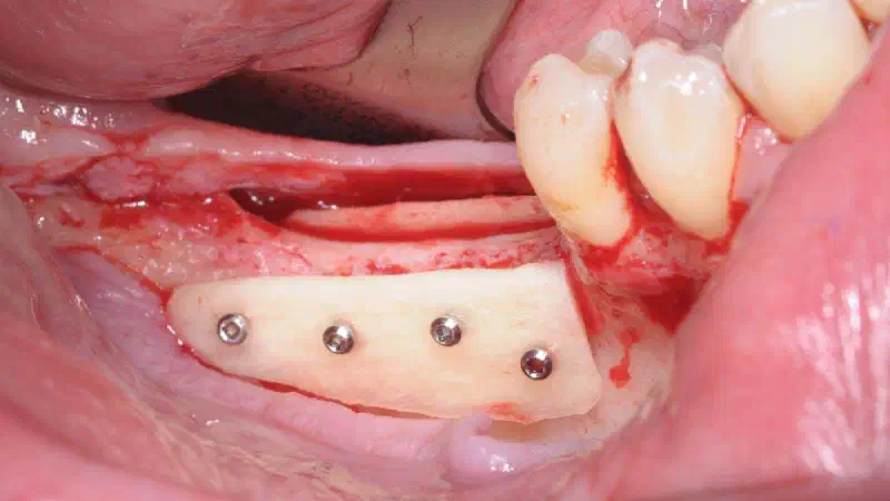
CASE REPORT Augmentation with allogenic and autogenous bone struts and consecutive relining in a split-mouth design (Original Title: Augmentation mit allogenen und autologen Knochenplatten und konsekutivem Relining im Split-Mouth Design) Tunkel J, de Stavola L. Zeitschrift für zahnärztliche Implantologie (Deutscher Ärzteverlag) 2019, 35, 2–11 In this case report, the shell technique with both autogenous and allogenic bone struts (maxgraft® cortico) was used for vertical bone augmentation in an atrophied mandible.
-
CASE REPORT Bone Augmentation and Simultaneous Implant Placement with Allogenic Bone Rings and Analysis of Its Purification Success https://www.mdpi.com/1996-1944/12/8/1291 Giesenhagen et al. (2019). Materials 2019, 12, 1291; doi:10.3390/ma12081291 CASE REPORT of #maxgraft® bonering shows successful regeneration of alveolar bone with a predictable clinical outcome, functionality and esthetics in different clinical indications. **Aim:**The main objective of this manuscript was to demonstrate the use of freeze-dried bone allografts (FDBA) by means of a technique of simultaneous bone augmentation and implant placement (“Bone Ring Technique”) in different indications, i.e., ridge reconstruction and sinus floor elevation procedure with a maxillary bone height of less than 4 mm.
-
CASE REPORT Case Report of maxgraft® bonering shows successful regeneration of buccal and lingual bone and immediate implant placement with a predictable clinical and esthetic outcome. Vertical bone augmentation in a single-tooth gap with an allogenic bone ring: Clinical considerations https://www.mdpi.com/1996-1944/12/8/1291 Giesenhagen et al. (2018). J Esthet Restor Dent. 2018;1–4 Objective: The main objective of this case report is to introduce a one-stage bone block augmentation with a cylindrical freeze-dried bone allograft (FDBA) and simultaneous implantation for the reconstruction of a single-tooth bone defect.
-
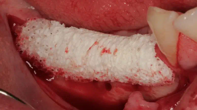
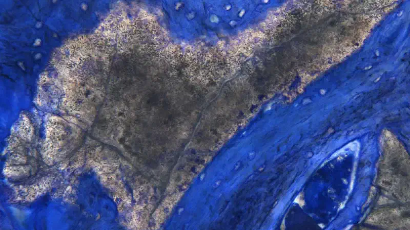
CASE REPORT Case report shows full remodelling of maxgraft® bonebuilder after 5 years Customized allogeneic bone grafts for maxillary horizontal augmentation: a 5 years follow-up radiographic and histologic evaluation Gruppe Kloss FR, Offermanns V, Donkiewicz P, Kloss-Brandstätter A. Clin Case Rep. 2020;00:1–8. https://onlinelibrary.wiley.com/doi/10.1002/ccr3.2777 This case report shows the full remodelling of maxgraft® bonebuilder after 5 years. Abstract We report the histological evaluation of an individualized allogeneic bone block 5 years after alveolar ridge augmentation. The biopsy showed a well-vascularized lamellar bone with fatty incorporations without any avital allograft remnants. The presence of osteocytes, lining cells, macrophages, and blood vessels indicated a healthy and vital bone tissue.
-
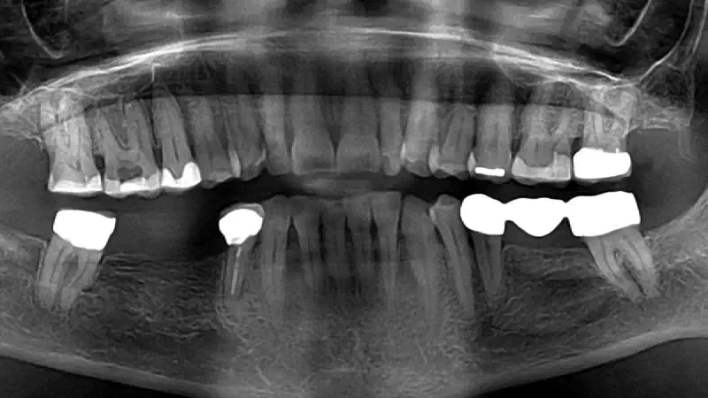
CASE REPORT Dental Implant Placement in Focal Osteoporotic Bone Marrow Defect: a Case Report and Treatment Recommendations Juodzbalys G. J Oral Maxillofac Res 2022;13(3):e5** http://www.ejomr.org/JOMR/archives/2022/3/e5/v13n3e5.pdf **doi: 10.5037/jomr.2022.13305 This case report provides insights into the management of a rare clinical condition mostly affecting the alveolar process of adult women and suggests a treatment protocol to achieve a successful implant-supported rehabilitation. CASE DESCRIPTION Panoramic x-ray of an edentulous site in the posterior lower jaw of a 54-year old woman revealed a large radiolucency with irregular borders, which was diagnosed a cortical focal osteoporotic bone marrow defect (FOBMD). These type of lesions are characterized by osteolytic hyperplasia of the hematopoietic bone marrow. As no clinical symptoms were present, an implant surgery was planned to bring back the missing dentition.
-
CASE REPORT Dreidimensionale allogene Knochenaugmentation in Schalentechnik Ein Fallbericht von Dr. Robert Würdinger In dem Fallbericht ist die Verwendung von allogenen Knochenplatten (maxgraft® cortico) in der Schalentechnik für eine komplexe dreidimensionale Knochenaugmentation beschrieben. Der Spaltraum wurde mit einer Mischung aus allogenen (maxgraft®) und autologen Knochenpartikeln aufgefüllt. Nach 4 Monaten konnte bei einer stabilen Knochen- und Weichgewebssituation implantiert werden. Bei fehlendem Knochenangebot zählt die Augmentation von Hartgewebe mit Knochenersatzmaterialien allogenen, autologen, xenogenen oder alloplastischen Ursprungs zu einem zentralen Erfolgsfaktor in der Implantologie. Allografts genießen bei dem Patienten eine inzwischen höhere Akzeptanz. Gegenüber den autologen Materialien bleibt dem Patienten bei dem Einsatz von Allografts eine Komorbidität der Entnahmestelle erspart und er wird dadurch weniger belastet. Begleitkomplikationen können bei der Entnahme verhindert werden, und darüber hinaus sind allogene Materialien unbegrenzt verfügbar. Der hier vorgestellte Fall beschreibt eine kombinierte horizontale und vertikale 3D-Knochenaugmentation in Schalentechnik mit allogenen Kortikalisplatten, allogener Spongiosa und aus der Umgebung gewonnenen autologen Knochenspänen.
-
CASE REPORT First Clinical Case Report on Alveolar Ridge Augmentation Using a Bovine Bone Substitute with Hyaluronate (cerabone® +HyA) Combined with Allogeneic Bone Granules (maxgraft®) Original title: First Clinical Case Report of a Xenograft–Allograft Combination for Alveolar Ridge Augmentation Using a Bovine Bone Substitute Material with Hyaluronate (Cerabone® +HyA) Combined with Allogeneic Bone Granules (Maxgraft®). Kloss, F.R.; Kämmerer, P.W.; Kloss-Brandstätter, A. J. Clin. Med. 2023, 12, 6214. https://doi.org/10.3390/jcm12196214 This case report demonstrates Guided Bone Regeneration for later implant placement in the aesthetic zone in a 34-year-old patient using a combination of freeze-dried bone allograft and pure bovine bone mineral with hyaluronate.
-
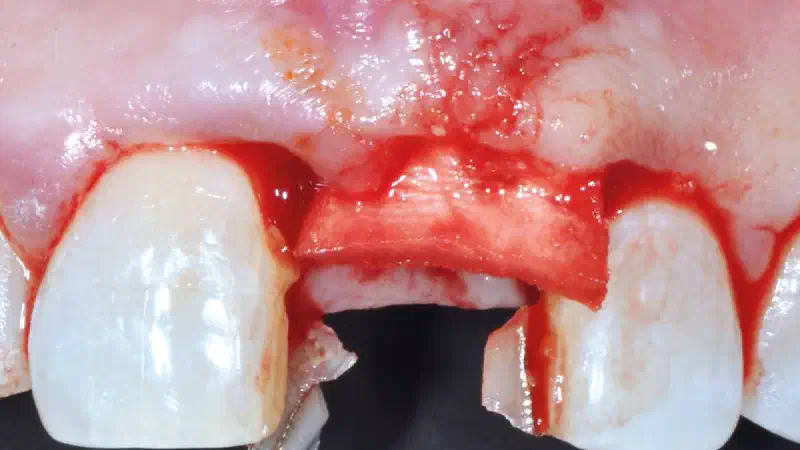
CASE REPORT Guided bone regeneration using layering technique in the aesthetic zone This case report describes the successful use of the layering technique with maxgraft® granules and cerabone® for guided bone regeneration (GBR) in the frontal maxilla. This technique was used to gain native bone around the implant and, at the same time, prevent resorption and obtain a positive aesthetic outcome in the long term through the slow resorbable properties of cerabone®. Initial situation A 68-year-old patient was referred from a colleague who already had to extract tooth #21 due to a failing post crown with cracked root. The patient is fit and healthy, not taking any medications, a non-smoker and with good oral hygiene. The missing front tooth site was left to heal for two months (Fig. 1). The occlusal view of the missing #21 showed severe buccal bone loss, which was even more visible after the flap was opened (Figs. 2-3).
-
CASE REPORT Guided Surgery for Bone Ring Technique A clinical case report by Vasilios Alevizakos, Ahmed Mohamed Othman, Constantin von See, Austria In totally and partially edentulous patients, implant placement and alveolar bone augmentation is a treatment that is considered to be very feasible and predictable in prosthetic rehabilitation1. Good clinical prognosis is due to the use not only of new materials but also of new technology, which aids the surgeon in providing the most accurate and precise treatment. The implant template is considered to be of clinical value, and can improve the accuracy of implantation and subsequently that of the overall implant solution2, 3. Different bone grafts are available depending on the amount of bone deficiency and the bone augmentation volume needed4-8. Bone grafts vary in nature, and can be autogenous, allogenic, xenogeneic, or alloplastic7. Allogenic bone grafts spare the patient from donor site morbidity and are known for their osteoconductive and osteoinductive properties.
-
CASE REPORT Guided Tissue Regeneration in Surgical Endodontic Treatment: Case Report and Literature Review Sidiropoulos, K., Roussou, K., Intzes, L., & Economides, N. (2019). Balkan Journal of Dental Medicine, 23(2), 102-107. https://content.sciendo.com/abstract/journals/bjdm/23/2/article-p102.xml This case report demonstrates the use of Guided Tissue Regeneration (GTR) for the treatment of a perio-endo lesion. Methods: Tooth 35 of a female patient was diagnosed with a pocket depth of 12 mm and grade two mobility. Radiographic analysis revealed an extended bone defect with involvement of the buccal plate and the presence of overextended filling material. Following root canal treatment, a periapical surgery using GTR was performed. After granulation tissue removal, cerabone® was applied, which was covered by a collagen membrane. The flap was closed and after one week, the sutures were removed.
-
CASE REPORT Hard tissue augmentation using an allogenic bone block: a clinical case report The high clinical survival rates has led to the fact that dental implants are often the treatment of choice for the treatment of single tooth gaps in the posterior region, where more minimally invasive and adhesive restorations are not indicated1. Unfortunately, severely atrophied alveolar crests are frequently observed after traumatic extraction of teeth, resulting in a lack of bone which may complicate the implant placement2. In such situations, an autologous bone block grafting procedure may be considered the golden standard of care, with high survival rates of both the bone block as well as the implants placed into it3.
-
CASE REPORT Horizontal ridge augmentation and implantation in the mandible using a Customized Allogenic Bone Block with BLX implants Orcan Yüksel Atrophy of the mandibular bone caused by premature tooth loss due to periodontal or endodontic problems can often be found in posterior areas. Problems associated with implantation in these cases often arise due to limited bone height or width of the mandible, and several treatment options have been suggested to gain enough bone for a stable implantation and achieve an esthetically good result.1 In clinical practice, freeze-dried bone allograft (FDBA) blocks have been used for alveolar ridge augmentation with promising results, offering patients a less-invasive treatment alternative to autogenous bone blocks, with no donor site morbidity and no second surgical site.2,3,4 Nowadays, customized allogenic bone blocks can be produced using the computer-aided design/computer-aided manufacturing (CAD/CAM) technology, enabling a shorter surgery time as manual block adjustment during surgery becomes unnecessary, thus enhancing patient comfort.5,6
-
CASE REPORT Ice Cream Cone Technique with permamem® Initial Situation A 66 year old healthy female patient presented at the dental office (no periodontal disease, non-smoker). On the 2nd of February 2021 a vertically fractured tooth number 2.5 (Figure 1) was diagnosed. A CBCT confirmed the clinical diagnosis. In addition, loss of the buccal plate was evident on the x-ray (Figure 2). Treatment Planning Based on the clinical diagnosis the decision was made to extract the unrestorable tooth and a socket preservation technique, i.e. Ice Cream Cone Technique (Elian et al. 2007), was proposed to the patient in order to recreate the missing bone and avoid the triangular aspect of the alveolus at the time of implant insertion.
-
CASE REPORT Immediate implant placement and function in the esthetic zone with a Straumann® BLX implant This case describes a successful treatment outcome of a hopeless tooth in the esthetic zone with thin gingival biotype using the Straumann® BLX Implant System, botiss cerabone® and botiss mucoderm®. Introduction Reaching and maintaining optimal gingival esthetics around implants in the anterior region is a challenging task. One of the main characteristics of immediate implant placement and provisionalization is its effectiveness in the esthetic outcome, preserving the existing bone and gingival architecture. Adequate primary stability is a prerequisite to enable this type of treatment. The implant design plays an important role in this context. According to recent clinical trials, the new Straumann® BLX implant offers the ideal properties for these clinical situations. At the same time, the SLActive® surface has an impact on the early osseointegration of immediately restored implants.
-
CASE REPORT Immediate placement and restoration of a new innovative fully-tapered implant replacing central and lateral incisors Dada K, Pariente L, Daas M, Montenero J. (2019) International Dentistry – African Edition Vol. 8, No. 6, p. 48 – 54. http://www.moderndentistrymedia.com/dec_jan2019/dada.pdf This case report describes the use and performance of a newly developed implant in immediate implant placement. Methods: Teeth 11 and 12 of a 52-year old patient were diagnosed hopeless and extracted consecutively. First, tooth 11 was extracted and an immediate implant placement procedure was performed using a Straumann® BLX Implant. Following implant insertion, a temporary abutment for the crown was installed and the gap between the implant and the buccal bone wall was augmented with cerabone® granulate. Finally, a screw-retained temporary crown was installed. Three months later tooth 12 was extracted and a socket preservation was performed using cerabone® and an autologous soft tissue punch. Two months following healing the final prosthetic restoration was done using a zirconia cantilevered implant supported bridge.
-
CASE REPORT Implant placement with simultaneous guided bone regeneration using 2mm healing abutment Algirdas Puisys, DDS, PhD, Viktorija Auzbikaviciute, DDS, Tomas Linkevicius DDS, PhD This case report shows successful implant placement with guided bone regeneration, simultaneous soft tissue thickening and submerged 2 mm healing abutment which we tend to call “decompression technique”. A particular technique was used in order to gain stable native bone around implant, prevent compression around implant neck and obtain proper space for the following prosthetic treatment with adequate emergence profile.
-
CASE REPORT Implant-prosthetic rehabilitation using individualized allogeneic bone blocks for bone defects caused by peri-implantitis Dr. Alice Josephine Müller, Simon Perez, Dr. Frank-Peter Strietzel Dtsch Zahnärztl Z Int 2020, 2, 8–13 https://www.online-dzz.com/archive/issue/article/dzzint-1-2020/5449-103238-dzz-int20200008-0013-implant-prosthetic-rehabilitation-using-individualized-allogenei/ This case report demonstrates the functional and esthetic reconstruction of a severe bone defect after peri-implantitis and periodontitis in the mandibular. **Introduction:**Implant-prosthetic rehabilitation after previous implant loss due to peri-implantitis and periodontitis is considered to be challenging and risky, as the remaining alveolar process defects are often extensive and may also affect neighboring regions. A 68-year-old female patient with advanced peri-implantitis in region 44 and a severely atrophied alveolar ridge in region 32–41 requested a new fixed implant-prosthetic restoration of the lower jaw. The patient had an inconspicuous general medical history and previous periodontitis therapy had been performed. Subsequent to explantation at site 44, pronounced horizontal and vertical defects of the alveolar ridge in region 44 to 46 and in region 32 to 41 were identified both clinically and using 3D radiographic imaging (cone-beam computed tomography, CBCT). Prior to implant planning, the status of peri-implantitis and periodontitis was re-evaluated, as there was a suspicion of residual cement-associated peri-implantitis, which could have resulted in implant loss.
-
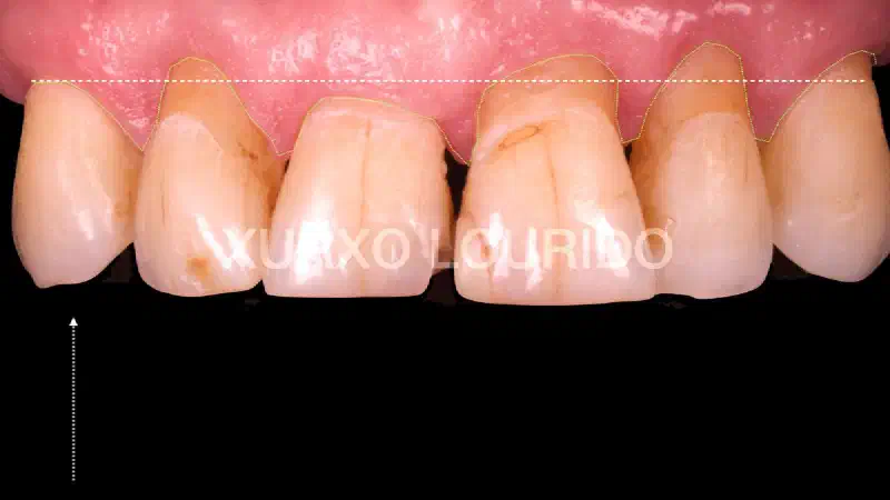
CASE REPORT Implant-supported rehabilitation of the maxillary ridge: Integration of surgical, periodontal and prosthetic concepts Xurxo Álvarez Lourido DDS. Dental technician Emilio Lopez Prado INITIAL SITUATION This case report describes the rehabilitation of the maxillary ridge, combining bone regeneration techniques and periodontal plastic surgery, together with novel techniques for the management of provisional prostheses, with the goal to return the patient a harmonious pink aesthetic outcome (Fürhauser et al. 2005).
-
CASE REPORT maxgraft® bonebuilder for alveolar reconstruction quantitated by a 3D matching technique: A case report https://doi.org/10.1002/ccr3.4771 Oliver Blume, Michael Back, Kim Martin, Péter Windisch**** First published: 12 September 2021 Original title: A customized allogenic bone block for alveolar reconstruction quantitated by a 3D matching technique: A case report Abstract An allograft, which was individualized into a precisely fitting bone block by CAD/CAM technology, was utilized for maxillary bone reconstruction. Six months after block insertion, an implant could be placed in an augmented alveolar ridge. The block’s volume and successful application were verified using a 3D matching software.
-
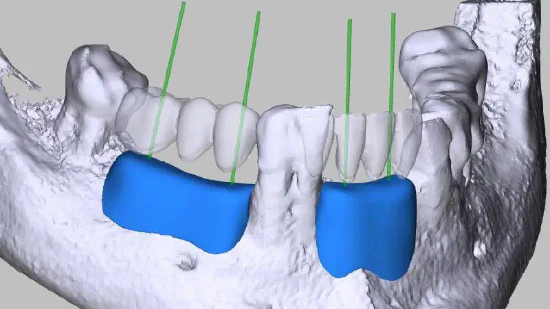
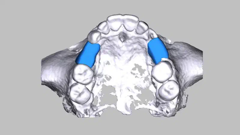
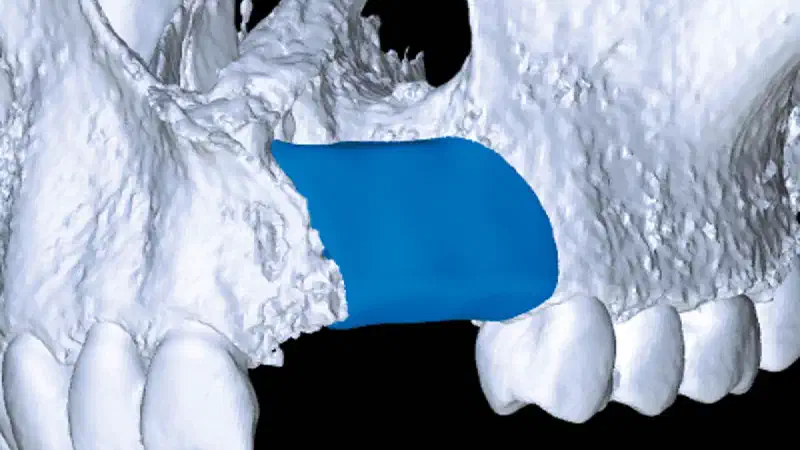
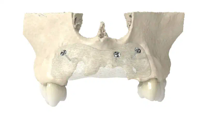
CASE REPORT maxgraft® bonebuilder for the regeneration of two large osseous defects in the maxilla Bilateral maxillary augmentation using CAD/CAM manufactured allogenic bone blocks for restoration of congenitally missing teeth: A case report Blume, P. Donkiewicz, M. Back, T. Born, J. Esthet. Restor. Dent. 2019, 1–8. DOI: 10.1111/jerd.12454 https://onlinelibrary.wiley.com/doi/abs/10.1111/jerd.12454 This case reports outlines the reconstruction of an extensive osseous defects with a customized allogenic bone block, maxgraft® bonebuilder. Clinical Considerations In this report, the regeneration of two large osseous defects in the maxilla with allogenic bone blocks made from human donor bone was demonstrated. The bone blocks were customized using the CAD/CAM technology in order to enable the insertion of four dental implants.
-
CASE REPORT Nasal Floor Elevation - An Option of Premaxilla Augmentation: A Case Report Jordan A, Vuletić M, Sušić M, Stojić L, Gabrić D. Surgeries. 2022; 3(4):306-313.**** https://doi.org/10.3390/surgeries3040033 The article presents a less invasive surgical technique for the reconstruction of the atrophic anterior upper jaw, with the goal to provide the patient an implant-supported denture and to increase quality of life. CASE DESCRIPTION The patient aimed for an alternative to his conventional removable denture in the maxilla. Assessment of the baseline situation by clinical and radiographic measures revealed extensively reduced bone volume in the upper jaw with pronounced pneumatization of the maxillary sinuses and shallow vestibule. Based on the patient anamnesis and consultation with a prosthodontist, augmentation of the missing bone employing nasal floor elevation and later placement of four implants as support for a bar-retained overdenture was planned.
-
CASE REPORT New indication for maxgraft® bonebuilder: First report worldwide about custom-milled allogeneic bone grafts for alveolar cleft osteoplasty Otto S, et al., Custom-milled individual allogeneic bone grafts for alveolar cleft osteoplasty – A technical note, Journal of Cranio-Maxillo-Facial Surgery (2017),Volume 45, Issue 12, December 2017, Pages 1955-1961 https://doi.org/10.1016/j.jcms.2017.09.011 Surgeons from the Ludwig-Maximilians-University in Munich, Germany, showcase in this technical note the successful repair of an alveolar cleft using maxgraft® bonebuilder, an individualized allogeneic bone graft, in a 36-year old female patient with a 6-month-follow up showing good bony integration.
-
CASE REPORT Plastic periodontal surgery combining mucoderm® and Emdogain® This case report describes the predictable coverage of multiple adjacent maxillary gingival recessions using the modified coronally advanced flap (MCAF) technique and modified coronally advanced tunnel technique (MCAT) in conjunction with an acellular porcine-derived collagen matrix (mucoderm®) combined with enamel matrix derivative (Straumann® Emdogain®). The author states: “In my opinion, this case can be viewed as satisfactory both for the patient and the operator, effectively demonstrating the benefits of the combination of mucoderm® and Emdogain® as regards time and pain.”
-
CASE REPORT Posterior mandible reconstruction using cerabone® +HyA, permamem® and mucoderm® – A case report The presented case describes the rehabilitation of a challenging extended edentulous site in the posterior lower jaw, using state-of the-art bone regeneration- and soft tissue management techniques, with the goal to bring back the patient a long-term stable dentition. INITIAL SITUATION A female patient (66 years old) came to the clinic requesting a periodontal treatment. The patient was in healthy condition, without taking medication and was a non-smoker. She was already missing tooth #36, #37 and #25 (Fig. 1 and 2). After initial periodontal treatment, revaluation and maintenance therapy, the patient requested to restore the missing teeth. The tooth gap at position #36 and #37 impaired the patient’s ability to chew and was therefore primarily of functional concern.
-
CASE REPORT Predictable horizontal ridge augmentation and soft tissue thickening using an allogenic bone strut and a porcine collagen membrane Sebastian Stavar Initial situation A 66-year old female, non-smoker in good general health, presented with missing teeth in sites 36, 45 and 46, which were extracted due to caries and fracturing. While the missing teeth in sites 36, 45 and 46 were initially restored with bridges, the patient wished for a fixed implant-supported prosthetic restoration in sites 45 and 46, due to a failure of the bridge. However, a pronounced loss of bone tissue in the posterior mandible (Cawood and Howell Class III), especially concerning the buccal wall, was observable at the initial assessment and further confirmed by radiographic imaging, so that direct implant placement was impossible (Figs. 1-3).5 The thin soft tissue in the region concerned posed an additional problem for proper treatment and aesthetic rehabilitation (Fig. 2).
-
CASE REPORT Reconstruction of a complex post-extraction defect using NOVAMag® membrane in combination with cerabone® and Jason® membrane Original title: Horizontal and Vertical Defect Management with a Novel Degradable Pure Magnesium Guided Bone Regeneration (GBR) Membrane—A Clinical Case Frosecchi, M. Horizontal and Vertical Defect Management with a Novel Degradable Pure Magnesium Guided Bone Regeneration (GBR) Membrane—A Clinical Case. Medicina 2023; 59(11): 2009. doi: 10.3390/medicina59112009 https://www.mdpi.com/1648-9144/59/11/2009 The presented case report highlights the application of NOVAMag® membrane for treating a complex defect with horizontal and vertical components. Following tooth extraction, the defect space was augmented with cerabone® and covered with the resorbable magnesium membrane to support and maintain space for the graft. Eventually, the whole augmentation site was covered with Jason® membrane and the flap was sutured for primary healing. During the follow-up period, there were no complications with the soft tissue. At 8 months, the re-entry was performed revealing good quality and quantity of bone with no remnants of the magnesium membrane. The edentulous area was successfully restored with an implant-supported bridge.
-
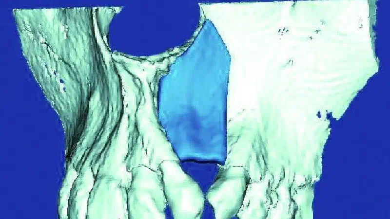
CASE REPORT Reconstruction of a Unilateral Alveolar Cleft Using a Customized Allogenic Bone Block and Subsequent Dental Implant Placement in an Adult Patient Blume O, Back M, Born T, Donkiewicz P. J. Oral Maxillofac. Surg., Article in press https://doi.org/10.1016/j.joms.2019.05.021 This case report presents the use of a customized allogenic bone block in a guided bone Regeneration (GBR) procedure for the reconstruction of a unilateral alveolar cleft and the successful insertion of dental implants in an adult patient.
-
CASE REPORT Reconstruction of the posterior maxilla using cerabone® +HyA – A case report Original title: Socket preservation, sinus lift and lateral augmentation by using natural bovine bone substitute with hyaluronate Zafiropoulos, G.; Trajkovski, B. Int J Dent Biomat Res 2023, 1, 1-6. https://ijdbr.com/index.php/ijdbr/article/view/12 CASE DESCRIPTION The patient presented with partial edentulism and pain in the in the posterior upper jaw. At this time point the region was supplied with a partial denture temporarily fixed on the teeth #13, #23, #27 and #28.
-
CASE REPORT Regeneration of Horizontal Bone Defect in Edentulous Maxilla Using the Allogenic Bone-Plate Shell Technique and a Composite Bone Graft - A Case Report **Kovac Z, Cabov T, Blaskovic M, Morelato L. Medicina (Kaunas). 2023 Mar 2;59(3):494. https://pubmed.ncbi.nlm.nih.gov/36984495/ ** This case report aimed to clinically and radiographically evaluate the efficacy of maxgraft® cortico combined with a composite bone graft (autologous bone/cerabone®) in the augmentation of a horizontal bone defect in the edentulous maxilla during a 6-year follow-up period.
-
CASE REPORT Replacement of a Failing Upper Anterior Bridge with Dental Implants INITIAL SITUATION A 75-year-old male patient developed a problem with a tooth supported bridge for the upper right central and left central and lateral incisor teeth that had been in situ for more than twenty years. One of the supporting teeth had become loose and the referring dentist noticed the upper left abutment tooth was grossly carious and the three-unit bridge entirely supported by the upper right central incisor tooth.
-
CASE REPORT Restoration of a 3D bony defect in region 46 with pre-fabricated allograft bone rings and simultaneous implantation A clinical case report by Bernhard Giesenhagen, Germany “The bone ring technique, as described in the following case report, has significant benefits over conventional procedures with autologous bone blocks: reduction in treatment time of between 45 and 60 minutes, avoidance of second procedure to harvest an autologous bone block and its associated risks, reduction of treatment time until the start of prosthetic restoration is reduced by three to six months.” (Dr. Bernhard Giesenhagen)
-
CASE REPORT Socket sealing with mucoderm® in the aesthetic zone Dr. Massimo Frosecchi This case report shows successful socket sealing technique after tooth extraction with a partially exposed mucoderm® of the upper central incisor in an aesthetically demanding case. Initial situation A 46-year old patient, non-smoker, ASA 1 and no periodontal disease presents with mild pain in the upper central incisor. Her central left incisor #21 showed a mobility and swelling. Additionally, the probing depth was 7-8 mm in buccal area. The preoperative radiographic control revealed a previous endodontically treated tooth with a fractured root.
-
CASE REPORT Three-dimensional augmentation with maxgraft® cortico A case report by Dr. Robert Würdinger In this case report, the usage of allogenic bone struts (maxgraft® cortico) in the shell technique for complex three-dimensional bone augmentation is demonstrated. The cavity between the struts and the alveolar ridge was filled with a mixture of allogenic cancellous (maxgraft® granules) and autologous bone particles obtained from the surrounding area. After 4 months healing period, it was possible to implant in a stable bone and soft tissue situation.
-
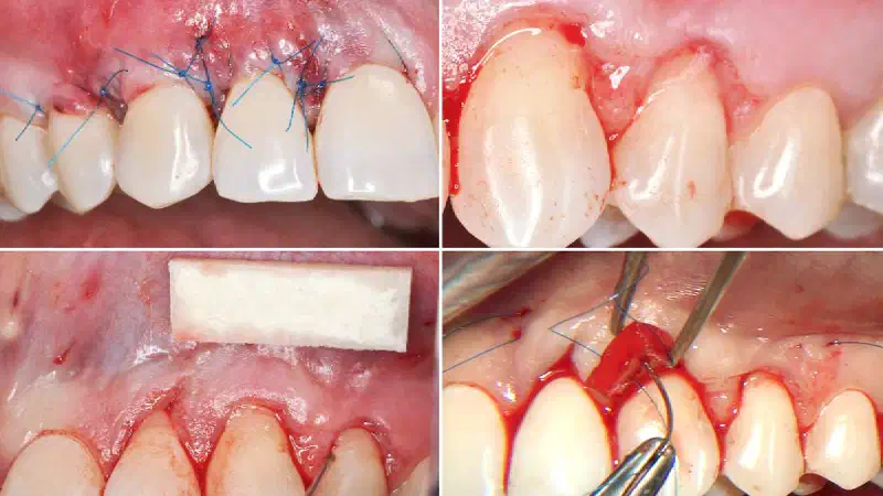
CASE REPORT Tips for optimized mucoderm® handling A clinical case report by Marcelo Nunes and Renata Cimões, Brazil This case report describes the success of mucoderm® in the treatment of generalized gingival recessions in a young patient. Initial situation A 24-year-old male patient without any medical history or smoking habits, well-controlled oral hygiene, presented generalized gingival recessions in upper jaw. The patient reported tooth brushing with high forces and no gentle tooth brush. Clinical examination evidenced Miller Class I recession without hypersensitivity.
-
CASE REPORT Treatment of Severely Resorbed Maxilla with maxgraft® bonebuilder Treatment of Severely Resorbed Maxilla Due to Peri-Implantitis by Guided Bone Regeneration Using a Customized Allogenic Bone Block materials-10-01213-v2-1 View Fullscreen Also interesting Study Efficacy and volume stability of maxgraft® bonebuilder – retrospective radiographic evaluation CLINICAL CASE 01/2021 Ridge augmentation with maxgraft® bonebuilder and vestibuloplasty with mucoderm® in the maxilla CASE REPORT **** Reconstruction of a Unilateral Alveolar Cleft Using a Customized Allogenic Bone Block and Subsequent Dental Implant Placement in an Adult Patient