

Complete restoration of a patient with advanced periimplantitis
Prof. Dr. Peter Windisch
Case_Peter_Windisch_210902_1_WEB-1
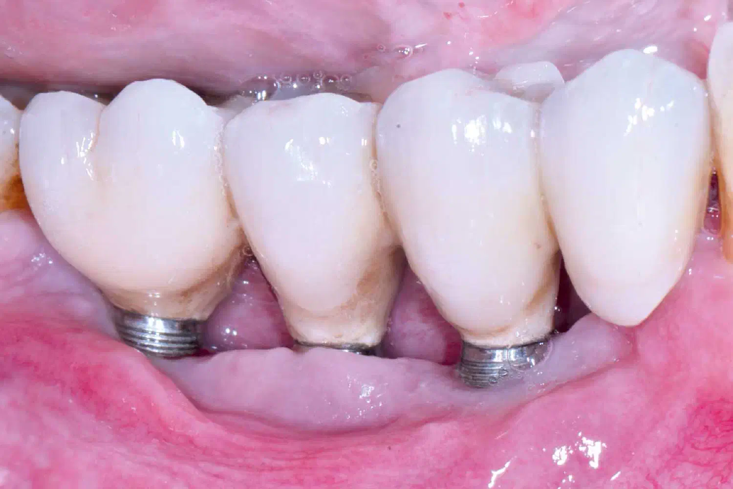
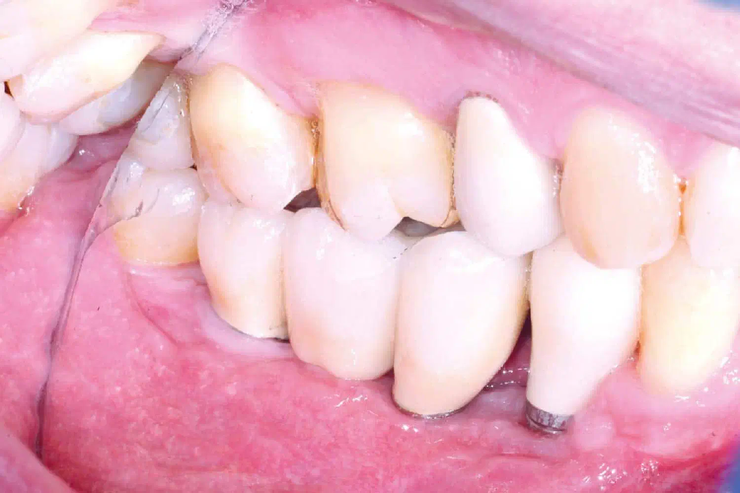
Initial situation: 3 ailing implants with a cantilever bridge construction.
Case_Peter_Windisch_210902_2_WEB-1
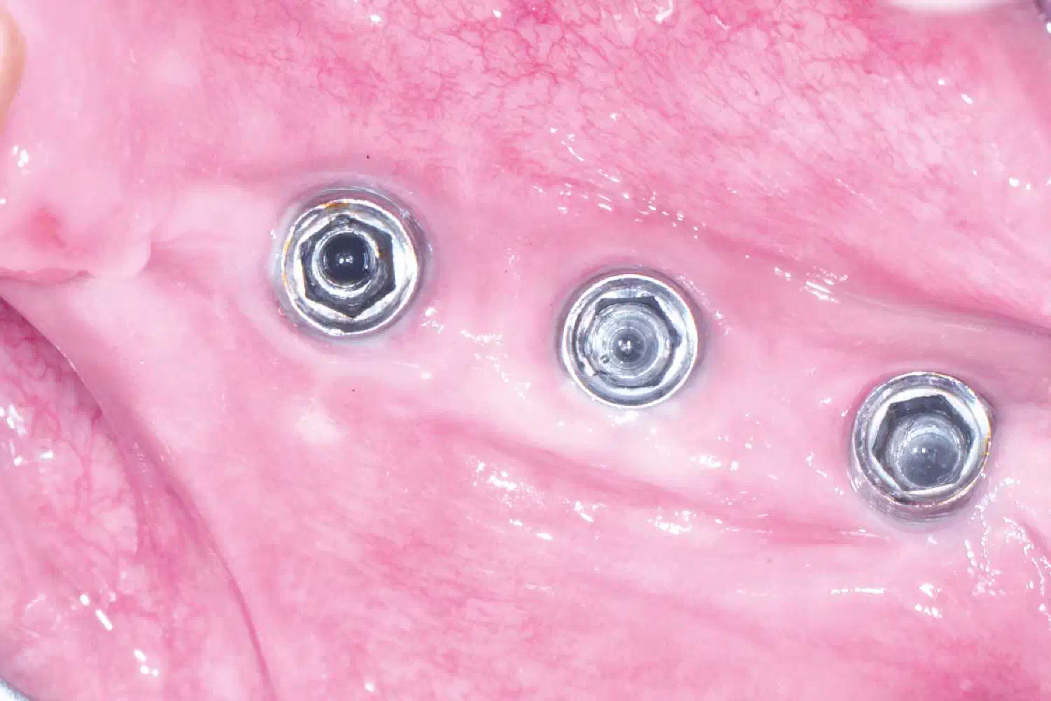
Occlusal view.
Case_Peter_Windisch_210902_3_WEB-1
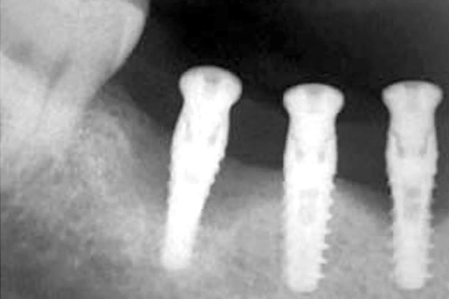
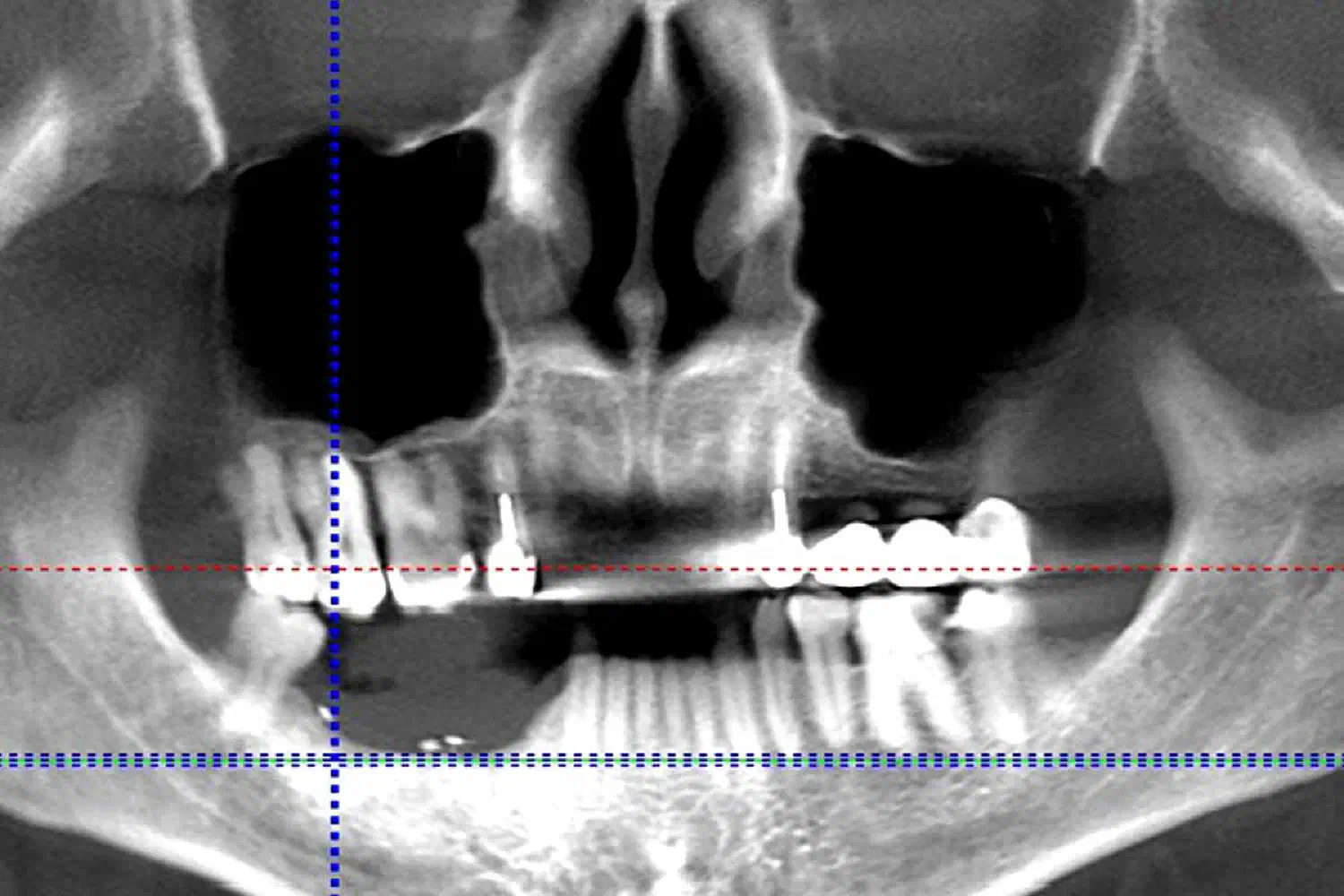
Radiographically detected periimplant bone loss.
Case_Peter_Windisch_210902_4_WEB
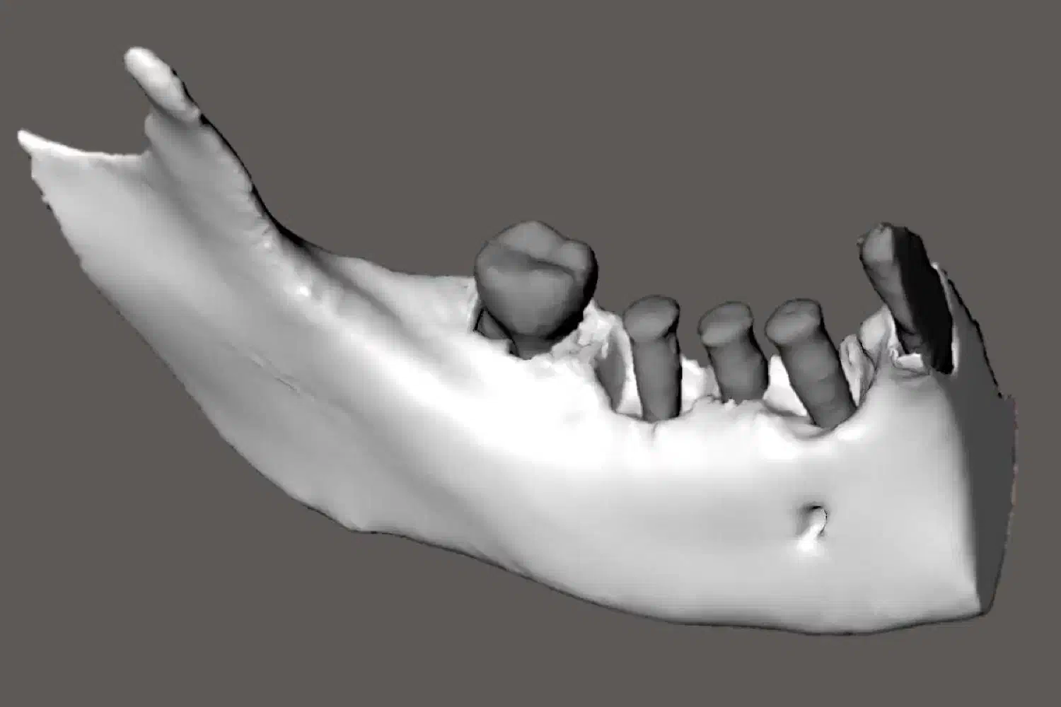
3D visualized periimplant defect morphology - digital CBCT evaluation.
Case_Peter_Windisch_210902_5_WEB-1
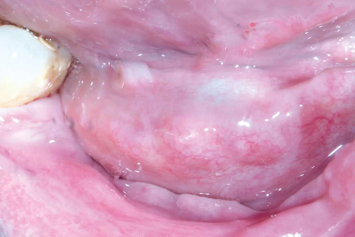
Situation after implant removal. The „superficial” (mostly affecting the soft tissue environment) inflammation was eliminated. Healing time 2-6 weeks.
Case_Peter_Windisch_210902_6_WEB-1
Situation after implant removal. The „superficial” (mostly affecting the soft tissue environment) inflammation was eliminated. Healing time 2-6 weeks.
Case_Peter_Windisch_210902_7_brb_WEB
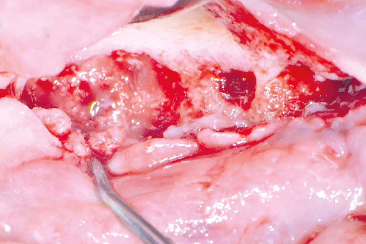
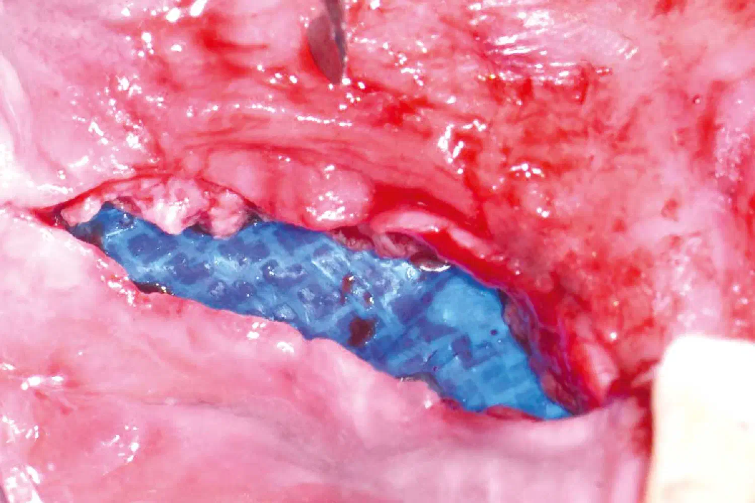
permamem® positioned above the advanced defect configuration. In addition, biotype modification was taken place using mucoderm®.
Case_Peter_Windisch_210902_8_brb_WEB
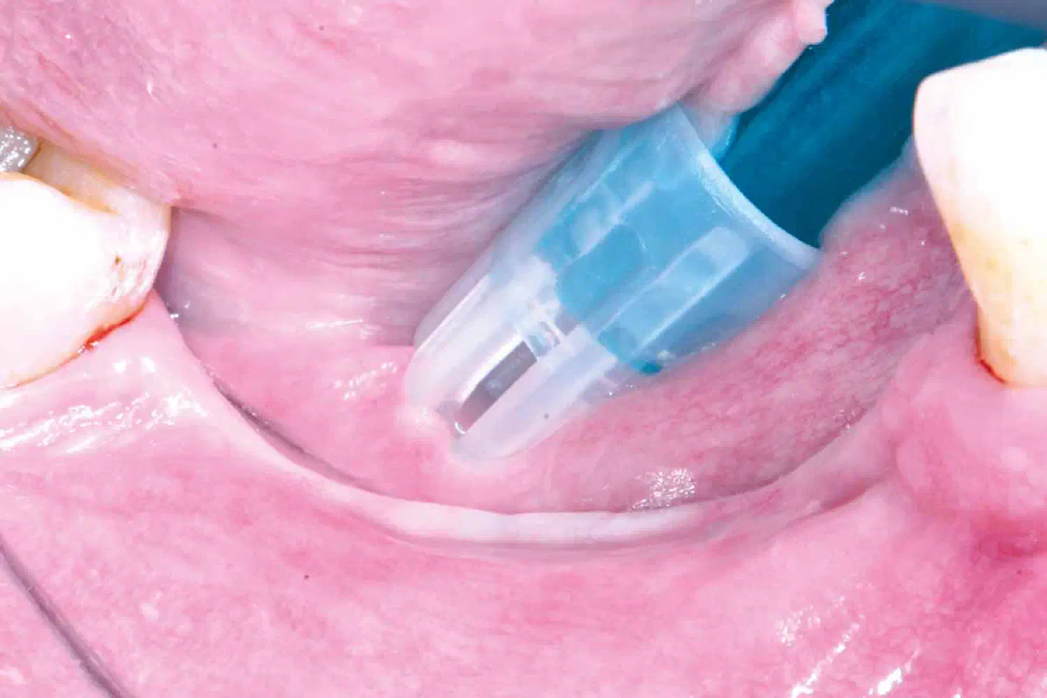
After extraction site development. Healing time 6 months.
Case_Peter_Windisch_210902_9_WEB-1
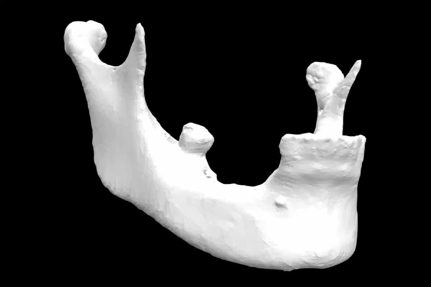
Volumetric changes of the hard tissue after extraction site development.
Case_Peter_Windisch_210902_10_WEB-1
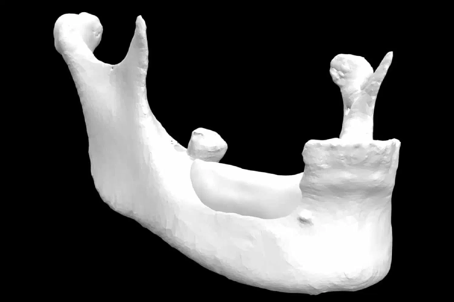
Planning for alveolar ridge augmentation
Case_Peter_Windisch_210902_11_WEB-1
Planning for alveolar ridge augmentation
Case_Peter_Windisch_210902_12_brb_WEB
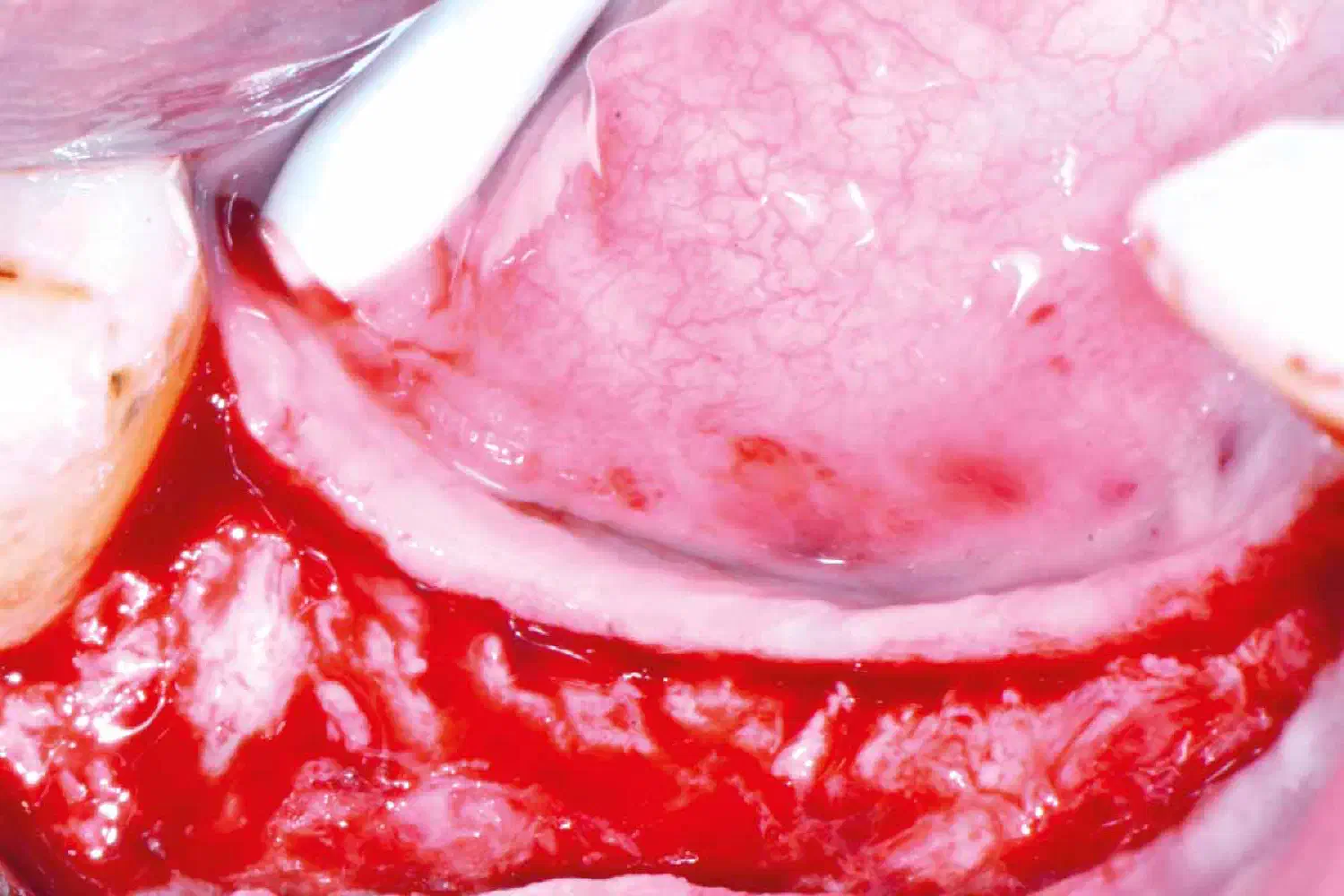
Second surgery for alveolar ridge augmentation.
Case_Peter_Windisch_210902_13_brb_WEB
Horizonto-vertical alveolar ridge augmentation with the application of permamem® above bone microblocks and composite graft consisting of 50% cerabone® and 50% locally harvested autogenous bone particles.
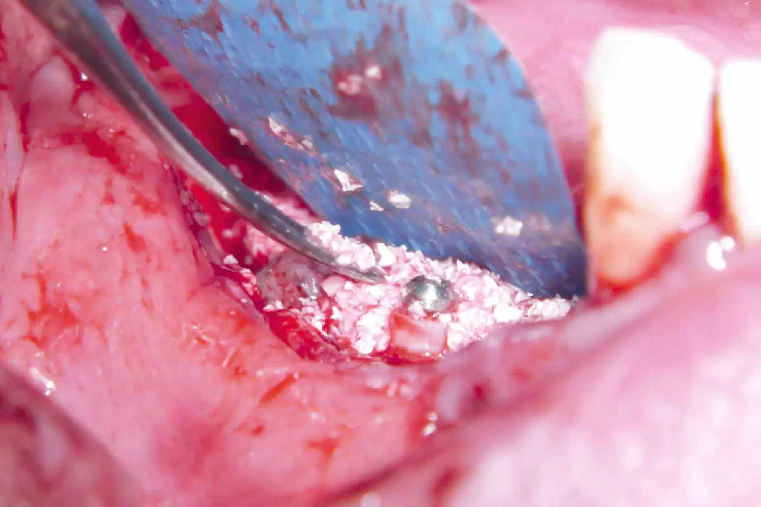
Case_Peter_Windisch_210902_14_brb_WEB-1
Situation before flap closure
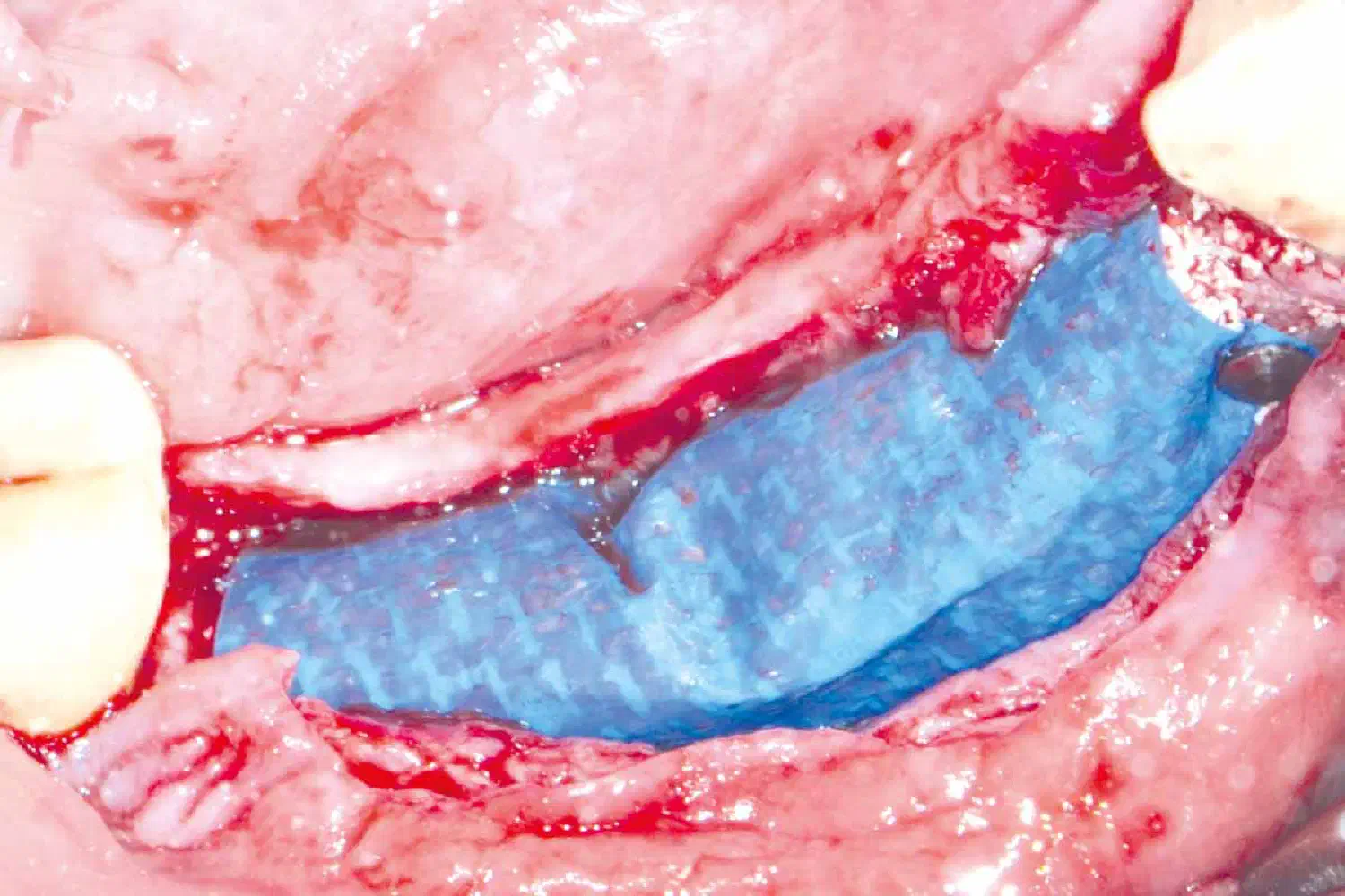
Case_Peter_Windisch_210902_15_brb_WEB-1
Early wound healing
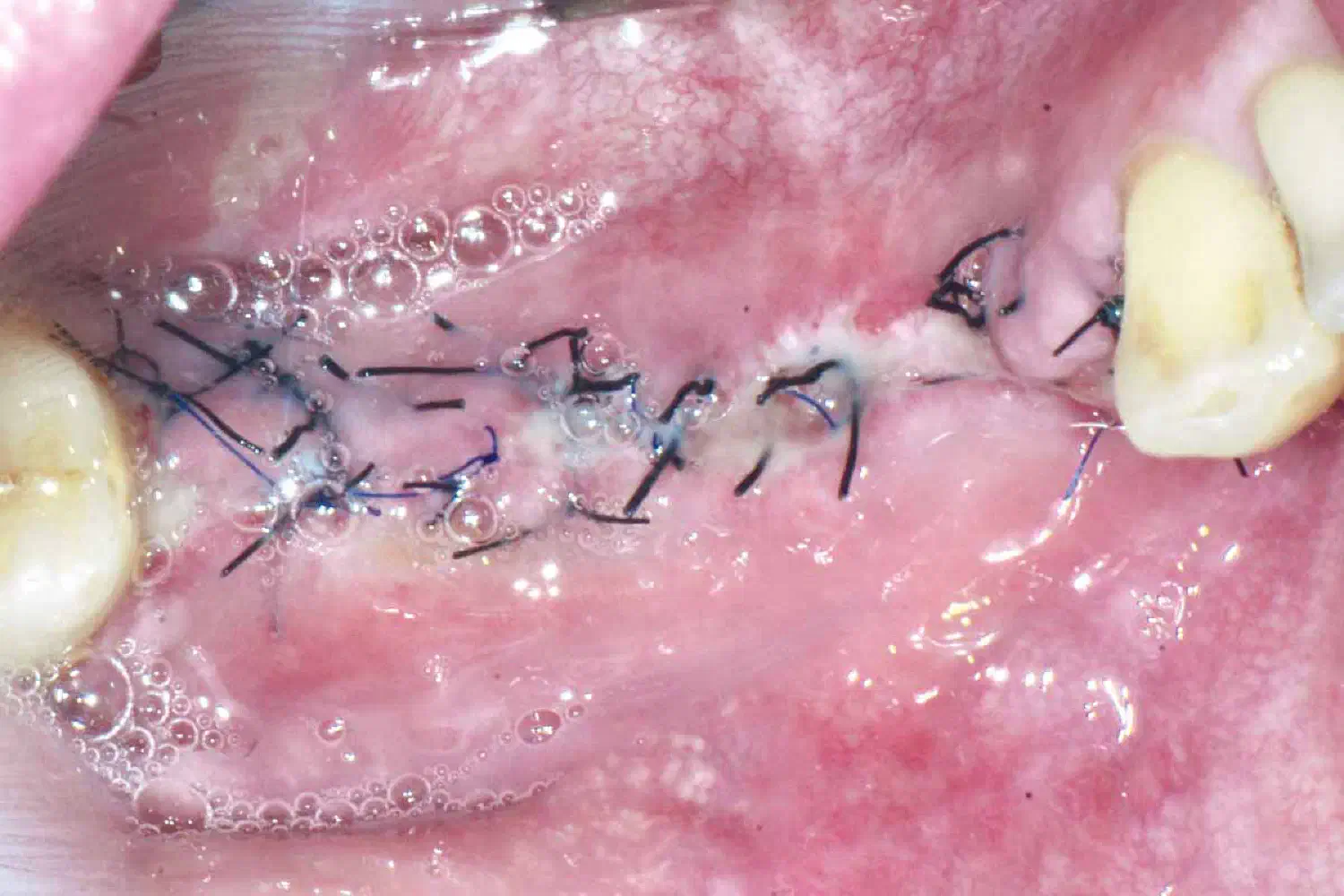
Case_Peter_Windisch_210902_16_WEB-1
Situation 6 months after alveolar ridge augmentation before re-entry. (3 months after augmentation a dehiscence occurred, therefore permamem® was removed and Jason® membrane was placed alongside collprotect® membrane).
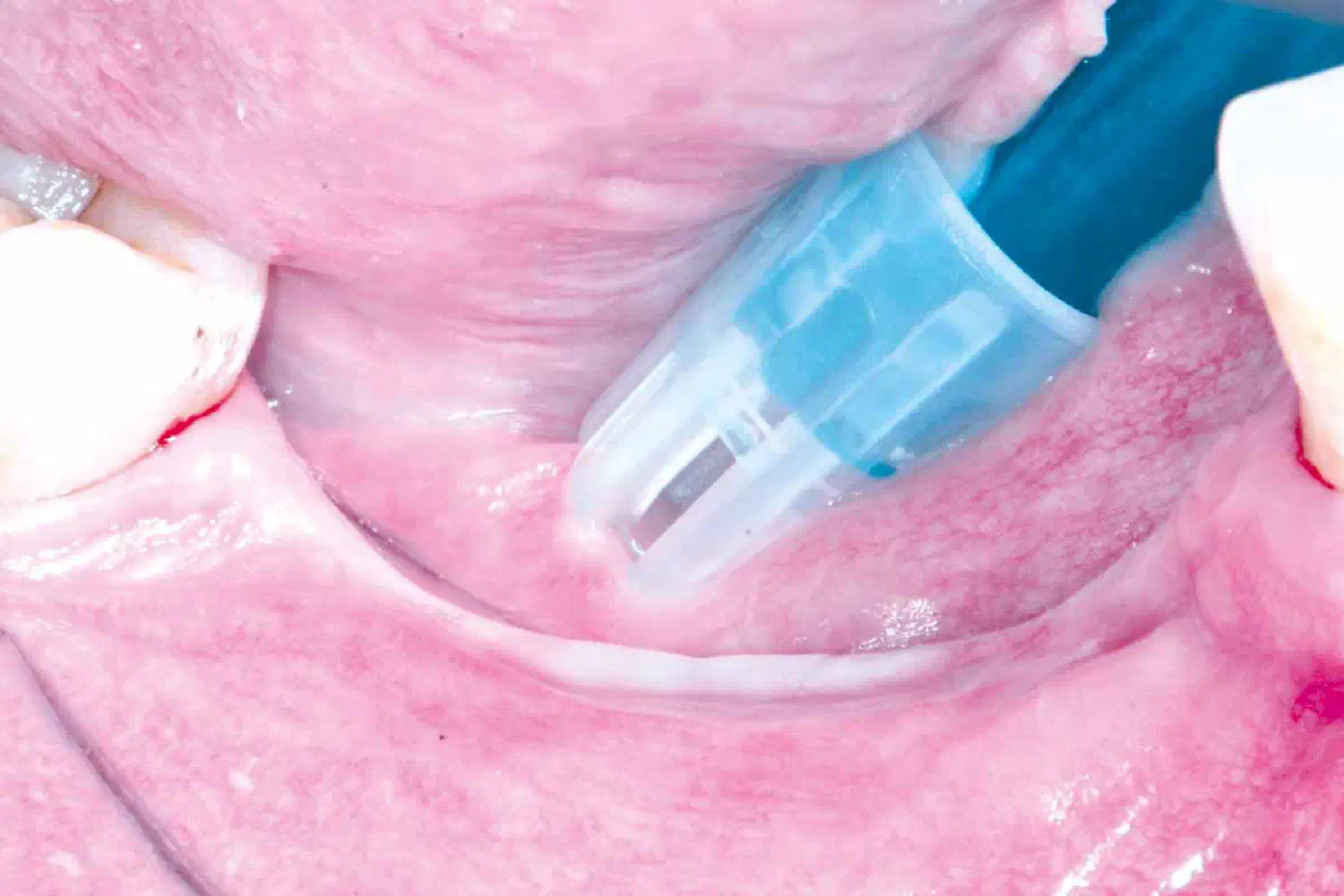
Case_Peter_Windisch_210902_17_WEB-1
Volumetric enhancement of the hard tissue.
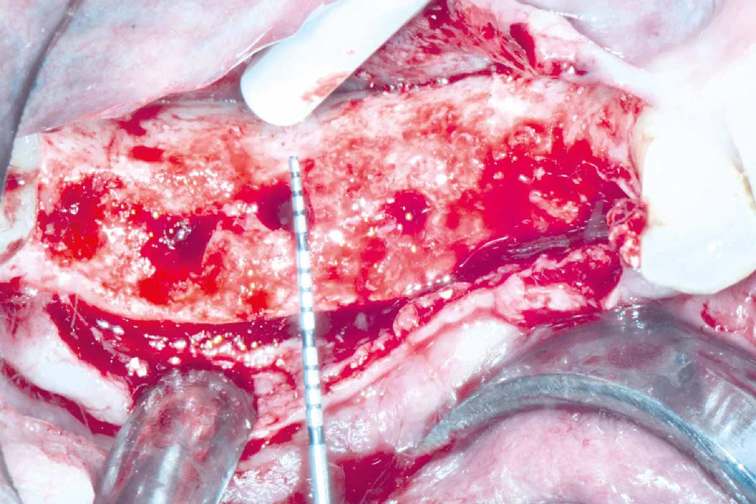
Case_Peter_Windisch_210902_18_brb_WEB-1
Implant placement in positions 44-45-47.
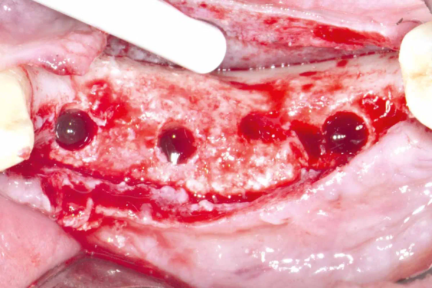
Case_Peter_Windisch_210902_19_WEB-1
Implant placement in positions 44-45-47.
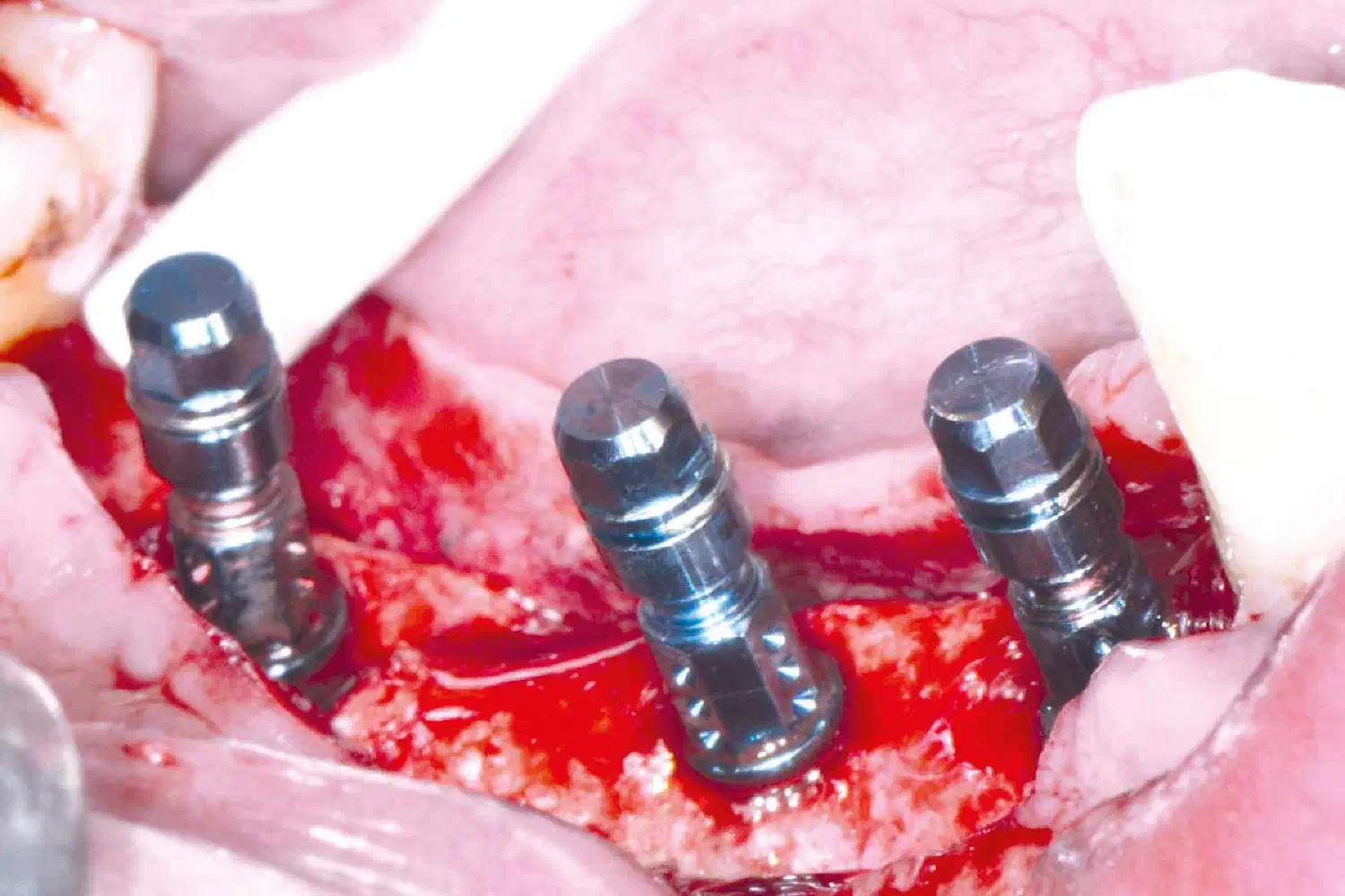
Case_Peter_Windisch_210902_20_brb_WEB-1
Simultaneous contour GBR using cerabone® mixed with autologous bone chips and collprotect® membrane.
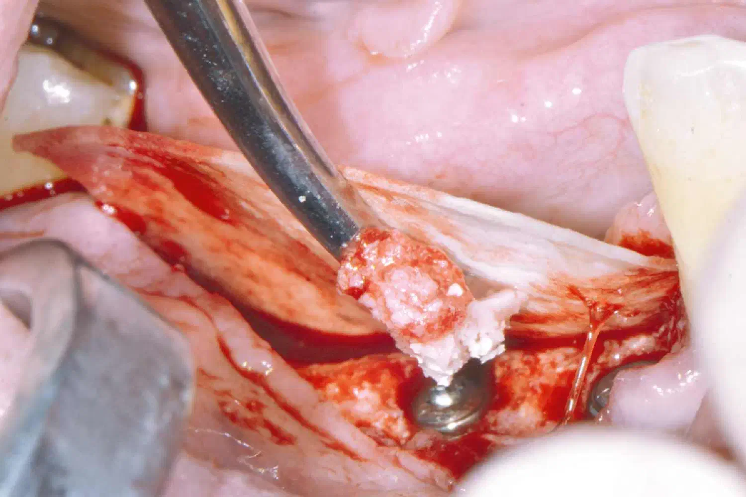
Case_Peter_Windisch_210902_21_brb_WEB-1
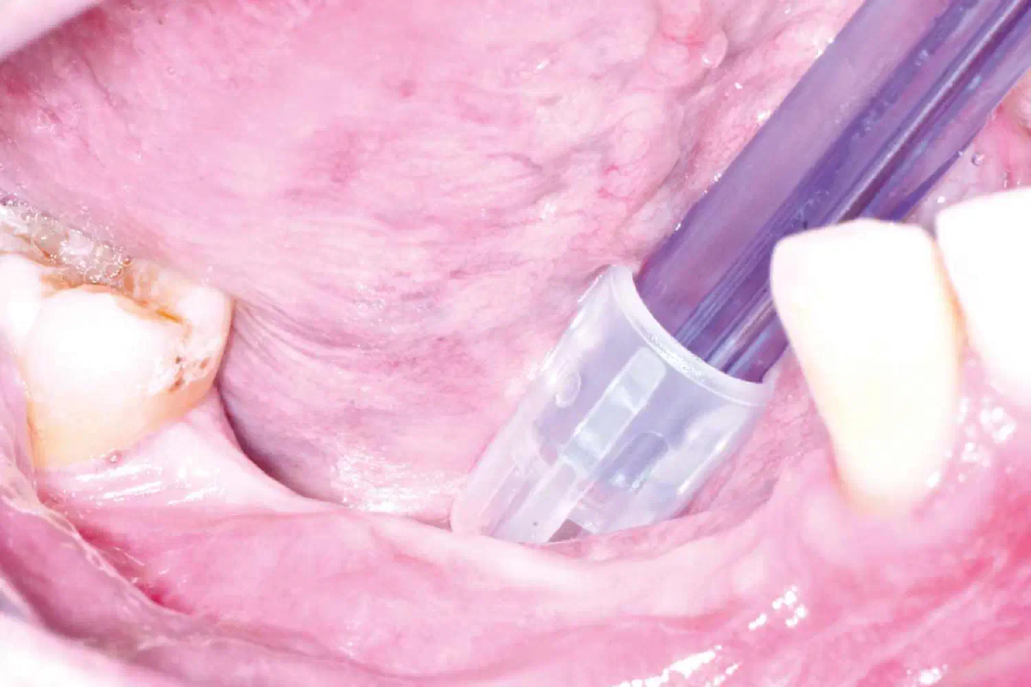
Situation 6 months following implant placement.
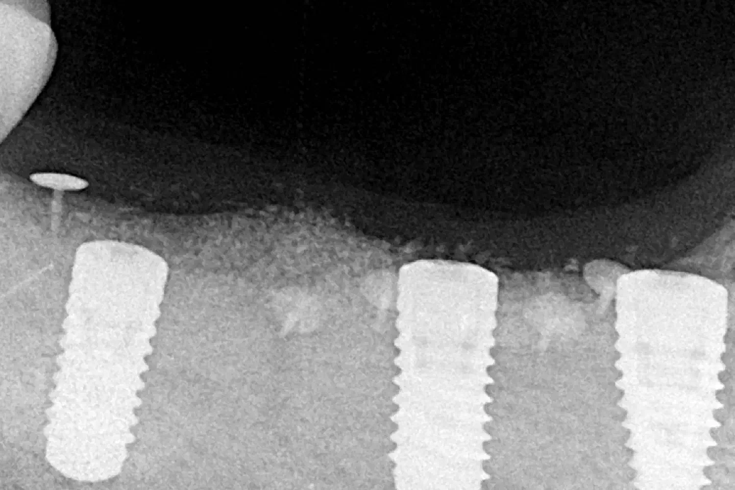
Case_Peter_Windisch_210902_22_WEB
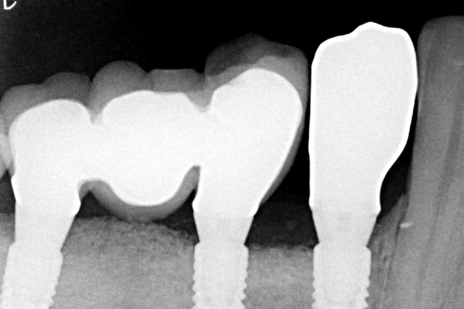
Radiographic control.
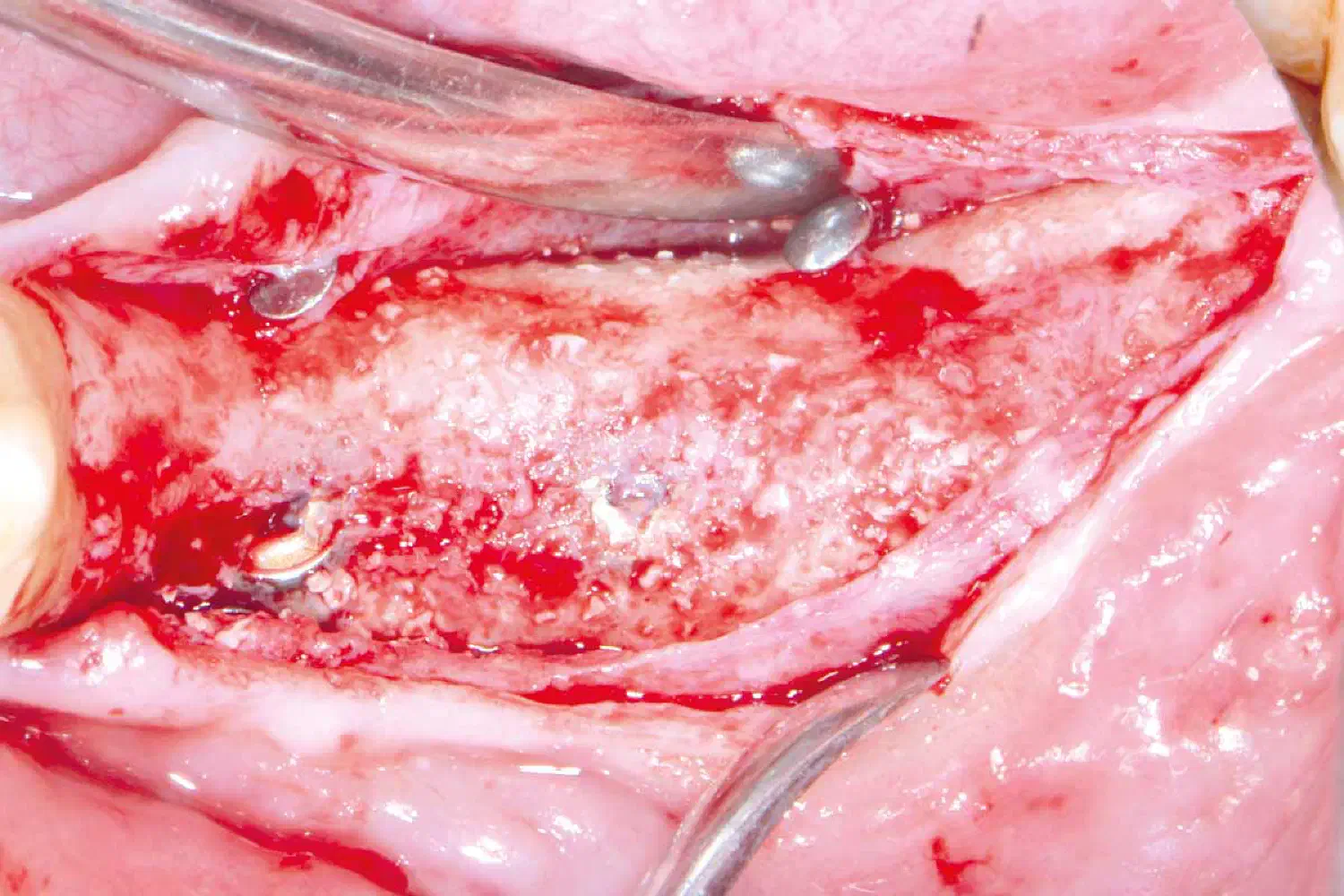
Case_Peter_Windisch_210902_23_brb_WEB-1
implant uncovering.
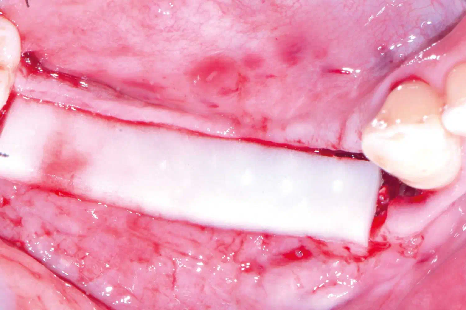
Case_Peter_Windisch_210902_24_WEB-1
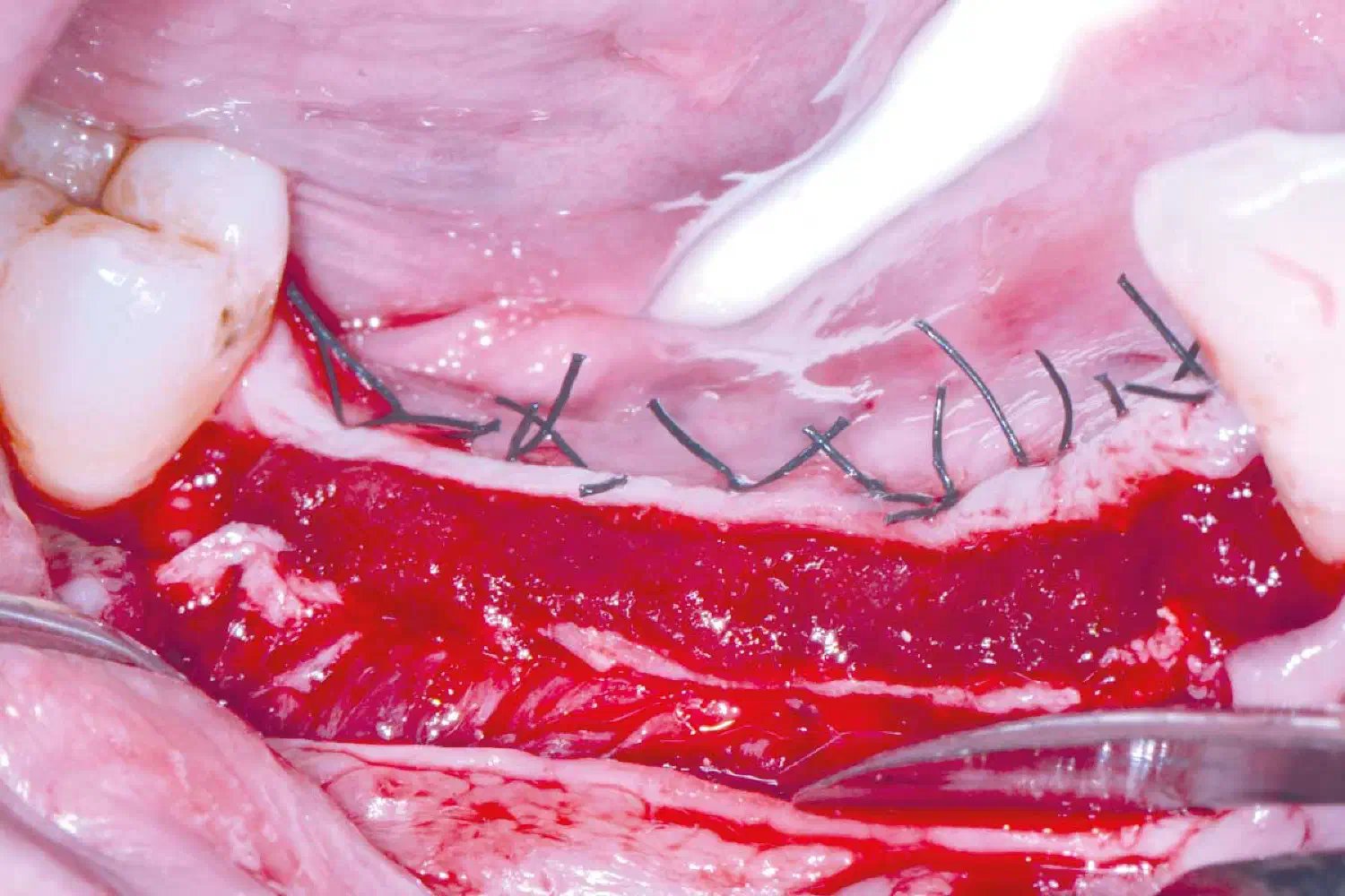
Soft tissue augmentation using mucoderm® to increase keratinized tissue thickness.
Case_Peter_Windisch_210902_25_WEB-1
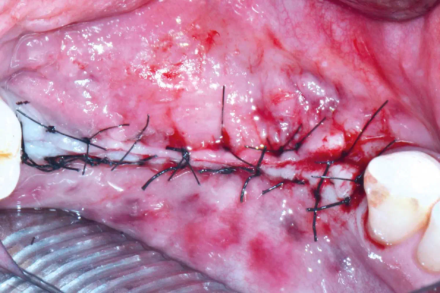
Two-layer wound closure.
Case_Peter_Windisch_210902_26_WEB-1
Two-layer wound closure.
Case_Peter_Windisch_210902_27_WEB-1
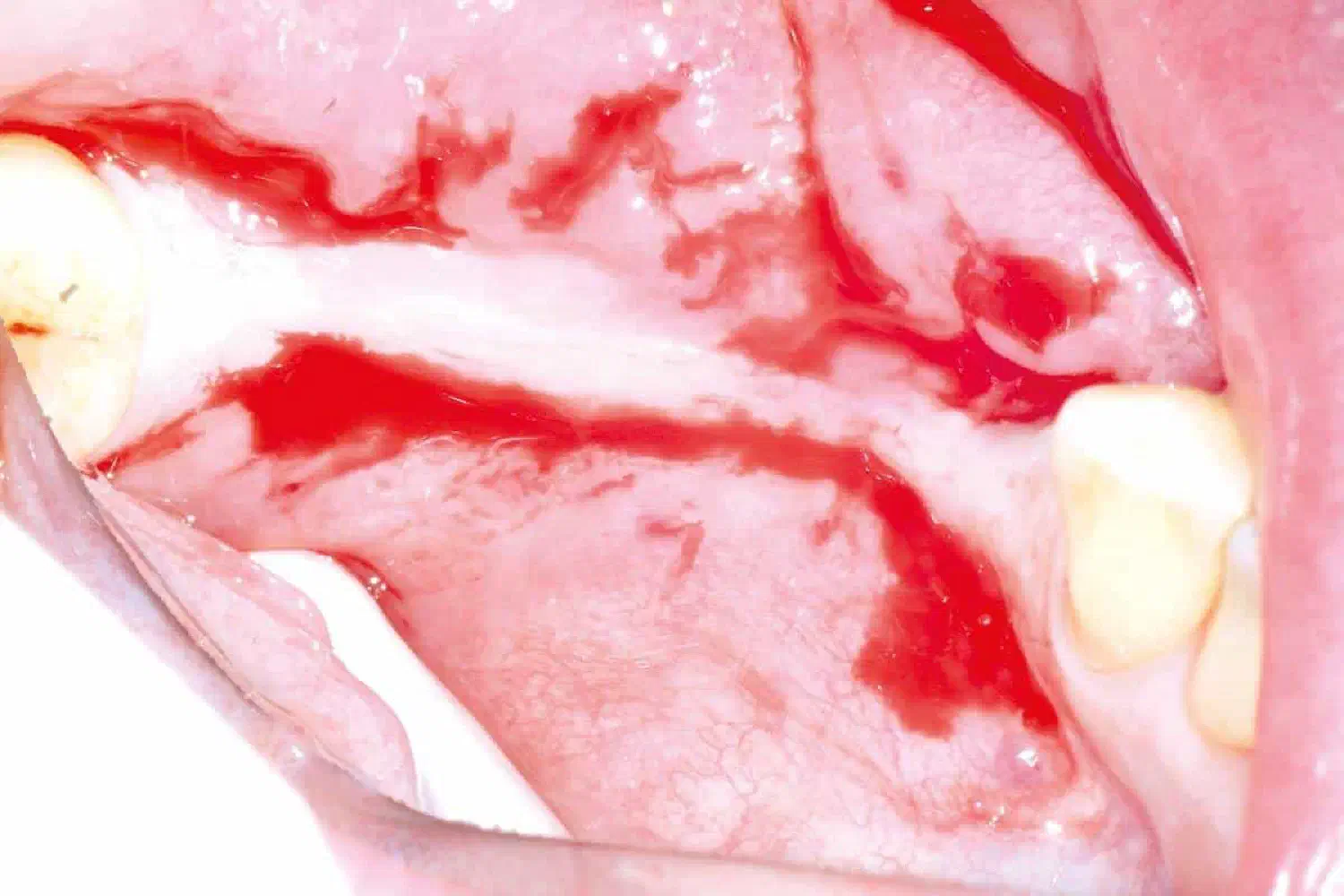
Situation 6 months after soft tissue augmentation.
Case_Peter_Windisch_210902_28_WEB-1
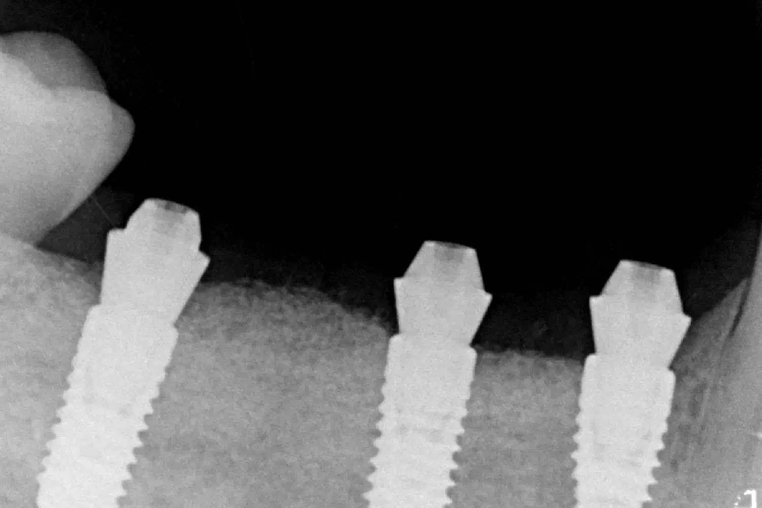
Abutment connection and final prosthetic rehabilitation: One-time abutments were secured at implant level and final prosthetic work was fabricated on abutment level, therefore dis- or reconnection could be avoided during the process.
Case_Peter_Windisch_210902_29_WEB-1
Final prosthetic work.
Case_Peter_Windisch_210902_30_WEB-1
Radiographic control.