

Staged implant placement following horizontal GBR with cerabone® +HyA, autologous bone, permamem® & Jason® membrane in the anterior maxilla
Dr. Joseph Gakonyo BDS, MSc
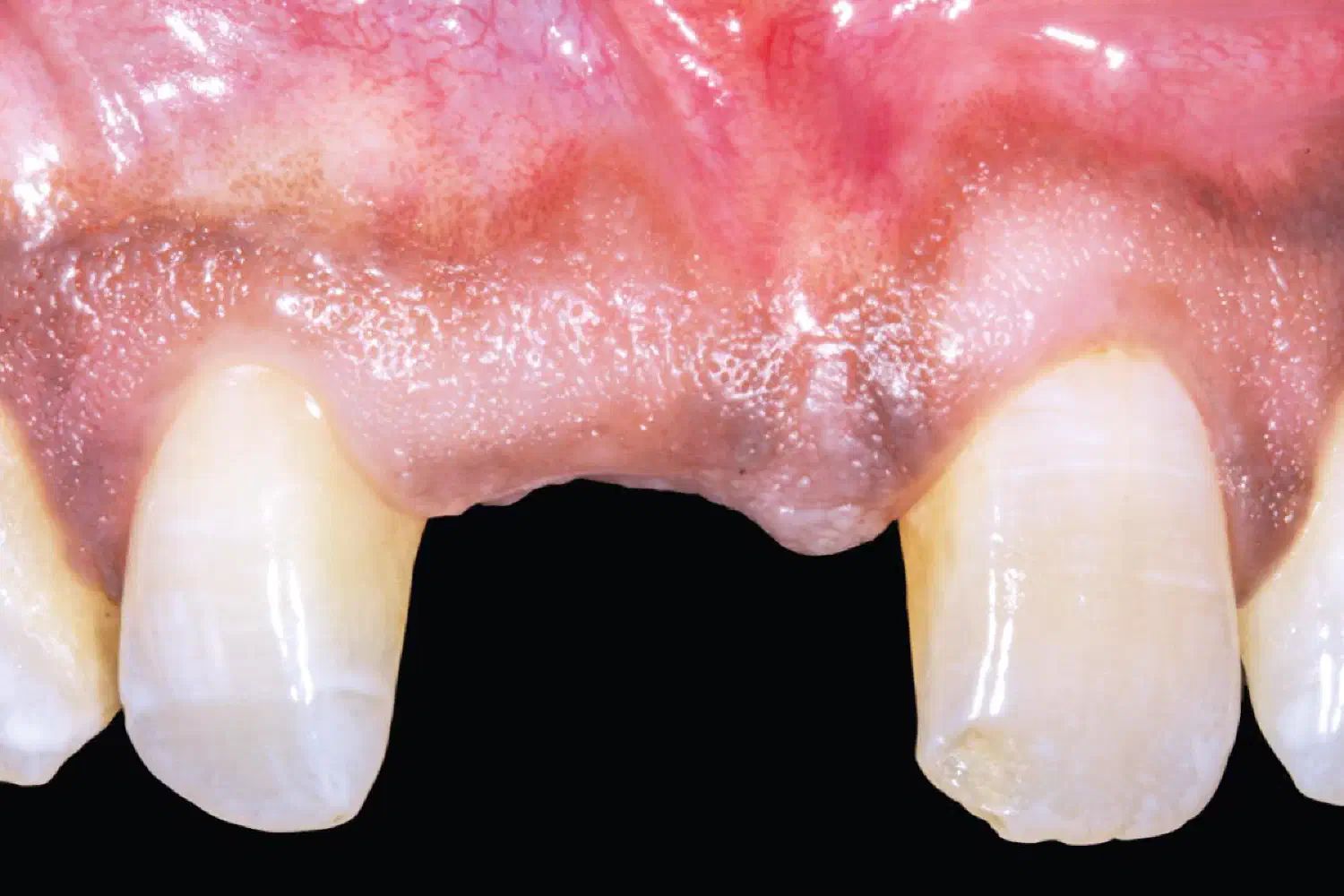
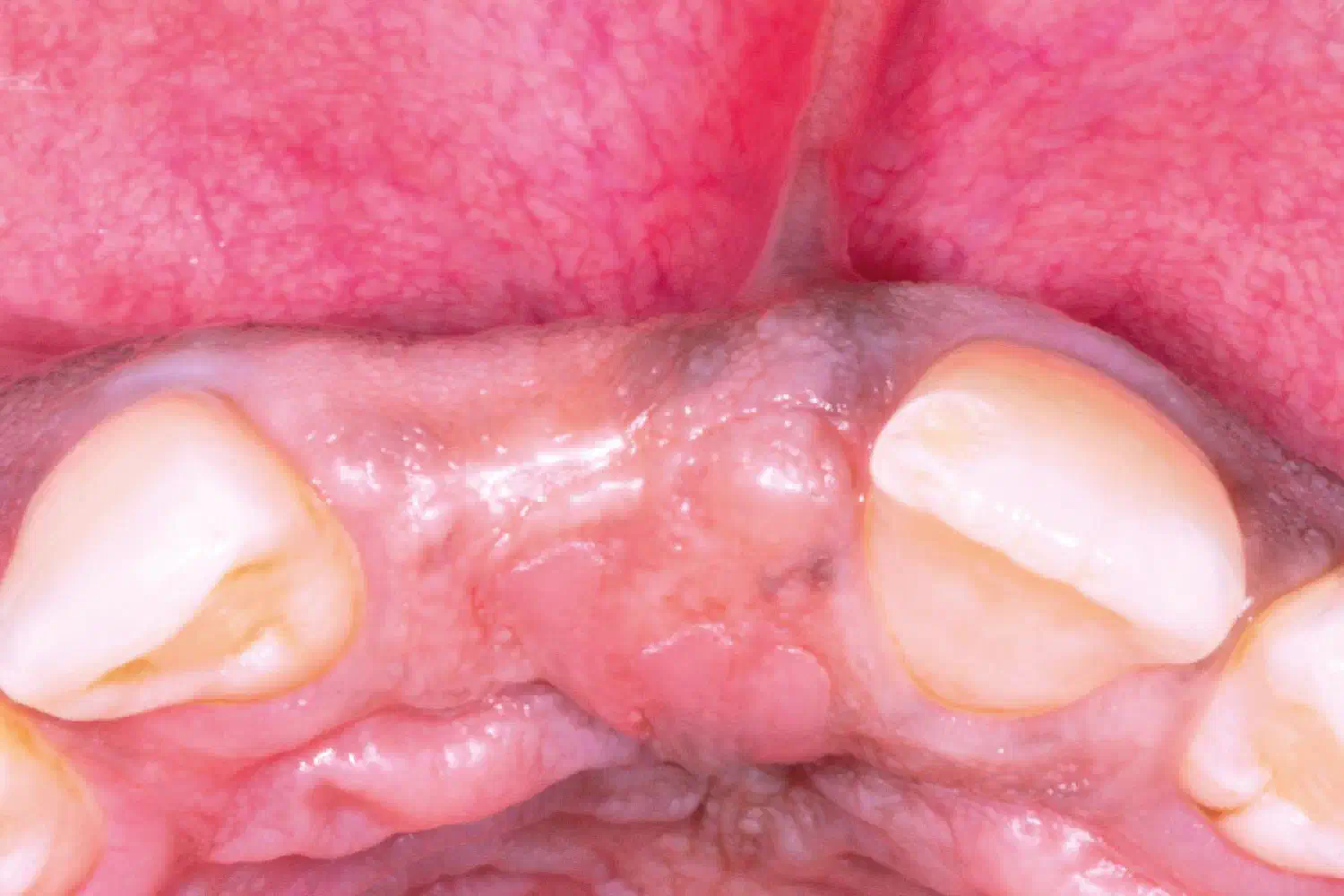
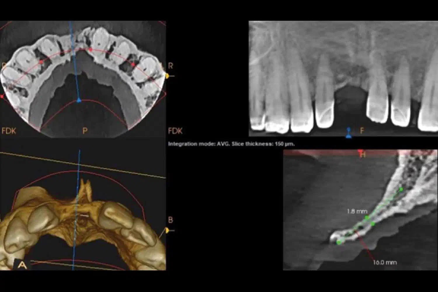
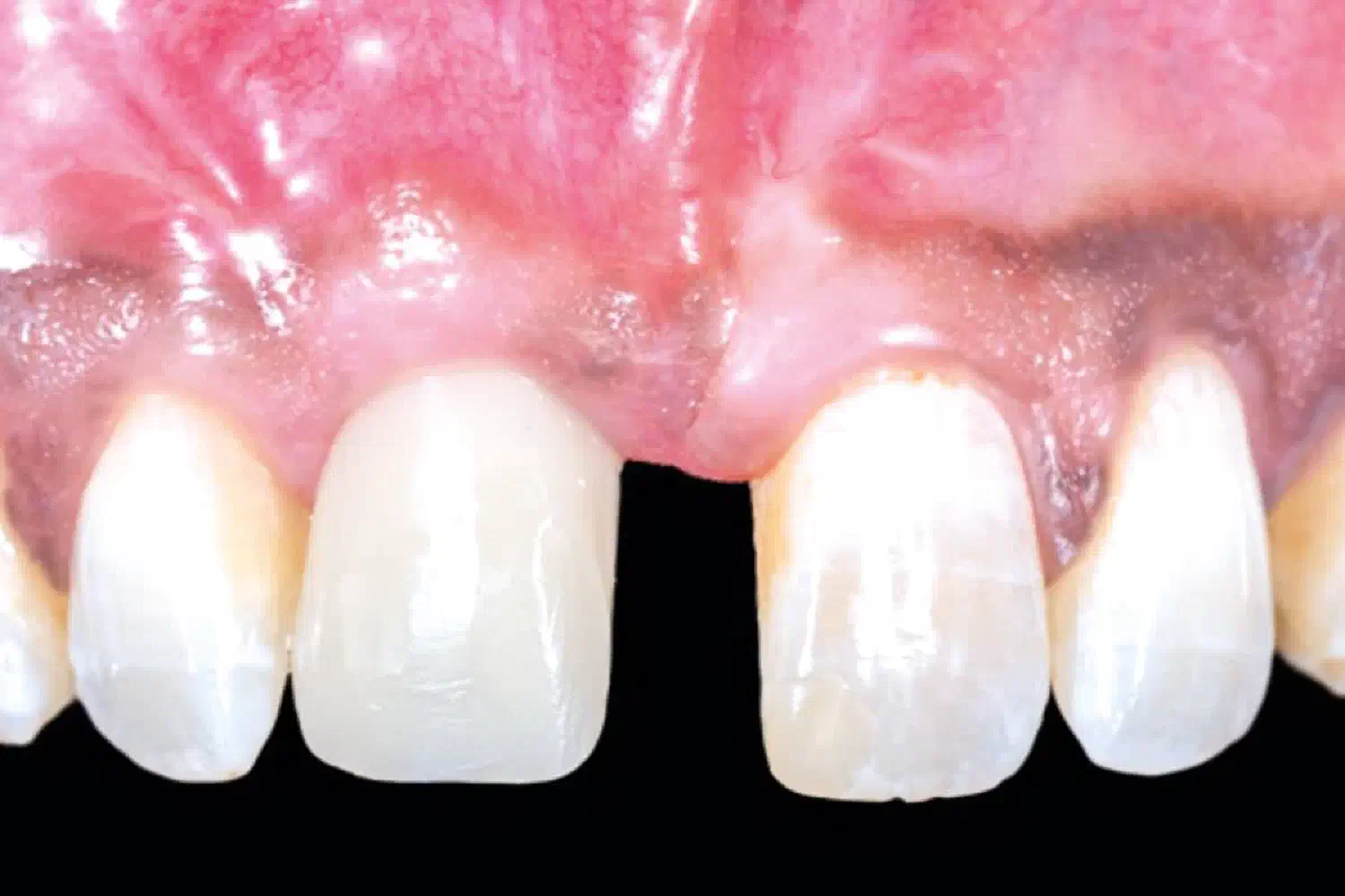
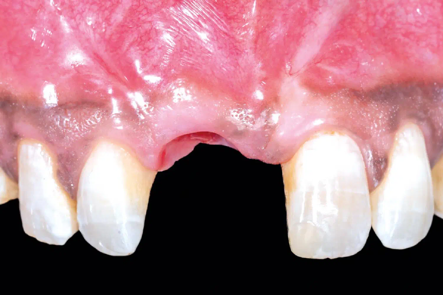
A healthy, non-smoking 48-year-old male patient visited our clinic due to the traumatic loss of his upper right central incisor at the age of 15. Upon clinical and radiographic assessment, it was determined that there was sufficient vertical bone but insufficient horizontal bone thickness (less than 2mm), which made simultaneous implantation with GBR unfeasible.
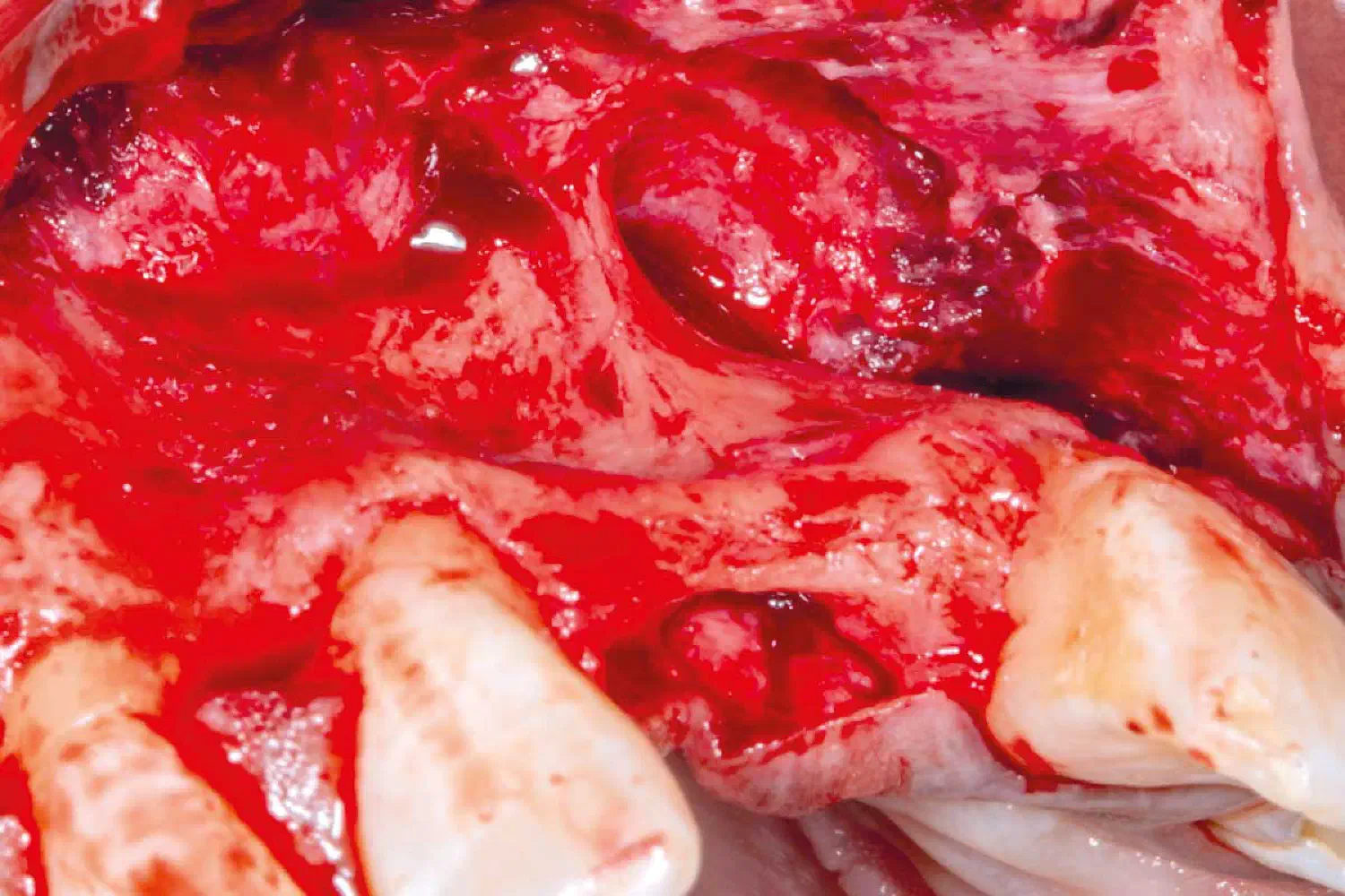
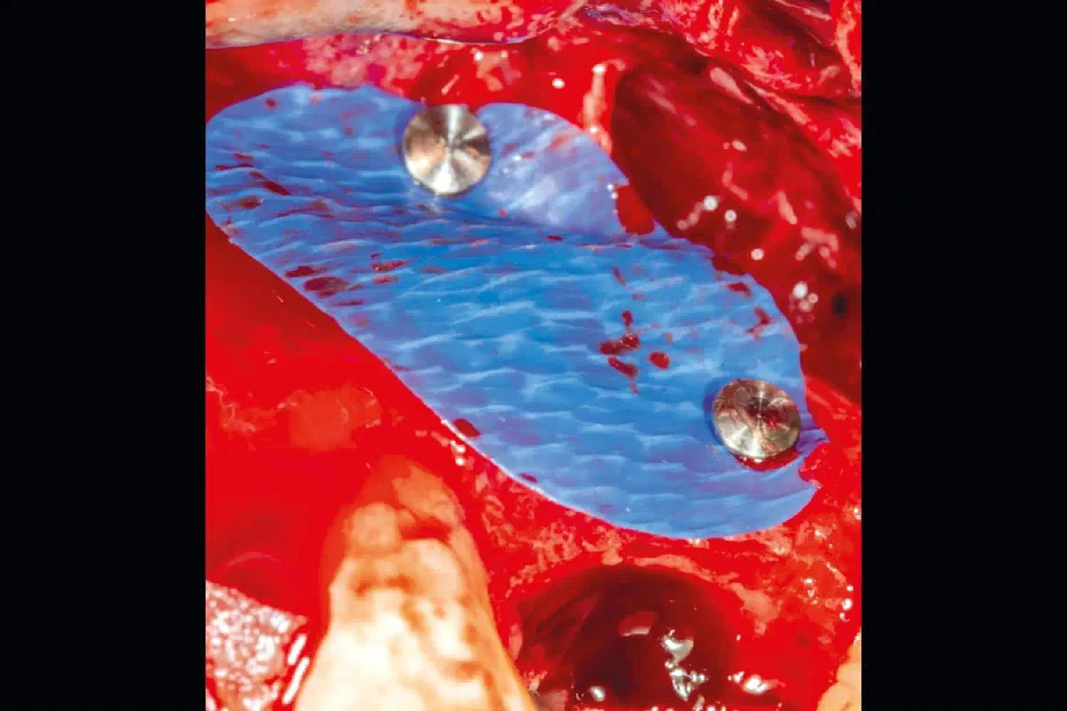
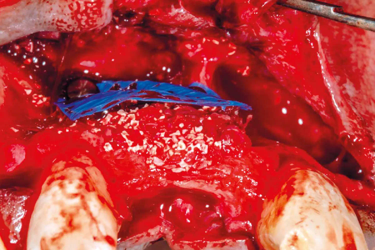
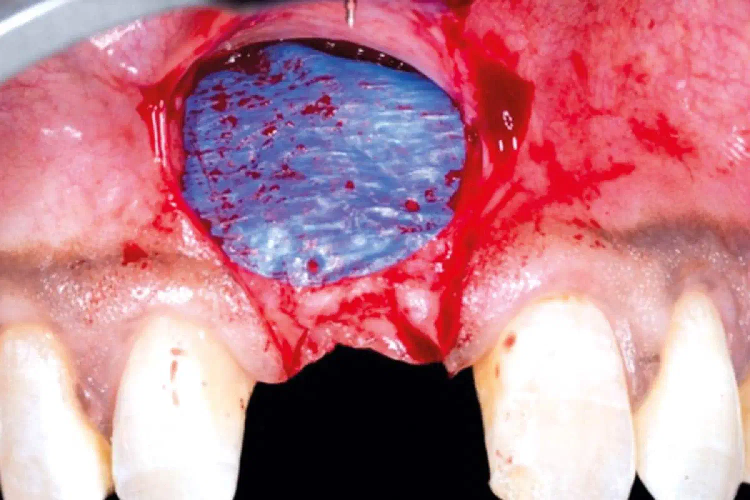
A guided bone regeneration procedure was carried out with autogenous bone mixed with cerabone® +HyA in a ratio of 70%:30% respectively. This mixed graft was covered using permamem® membrane fixed to the local bone using titan pins . Following, Jason® membrane was used to cover the non-resorbable PTFE membrane to protect the overlying mucoperiosteal flap. A de-epithelialized connective tissue graft harvested from posterior palate was fixed to the inner aspect of the mucoperiosteal flap to increase soft tissue thickness buccal and crestal to the future implant site.
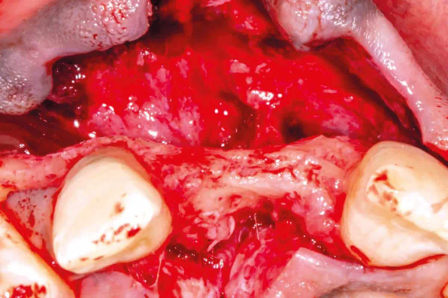
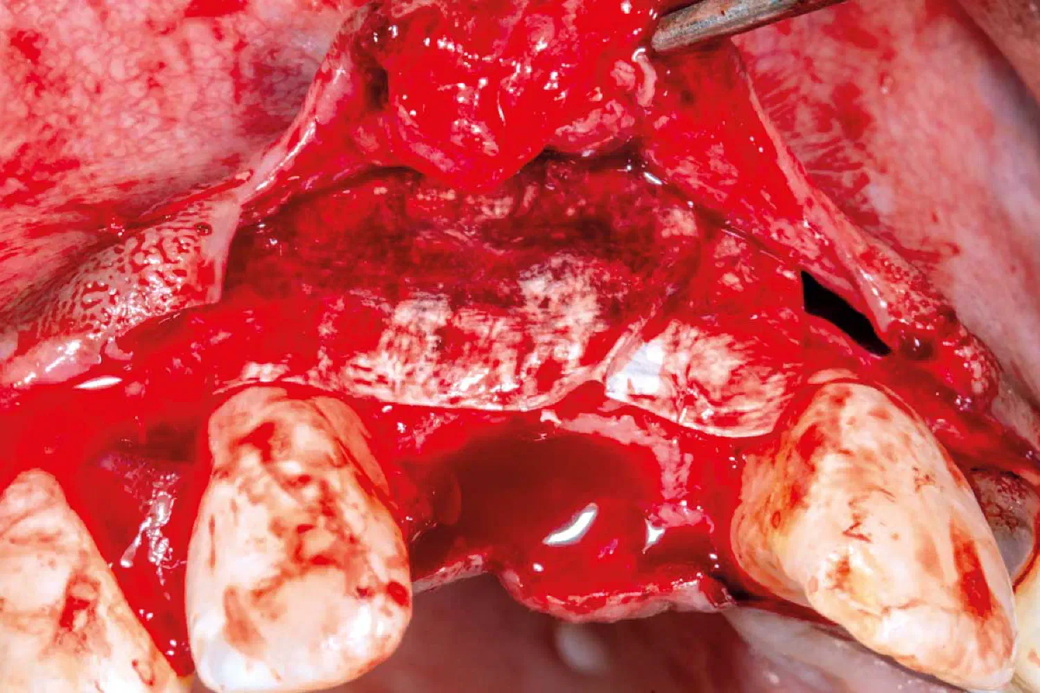
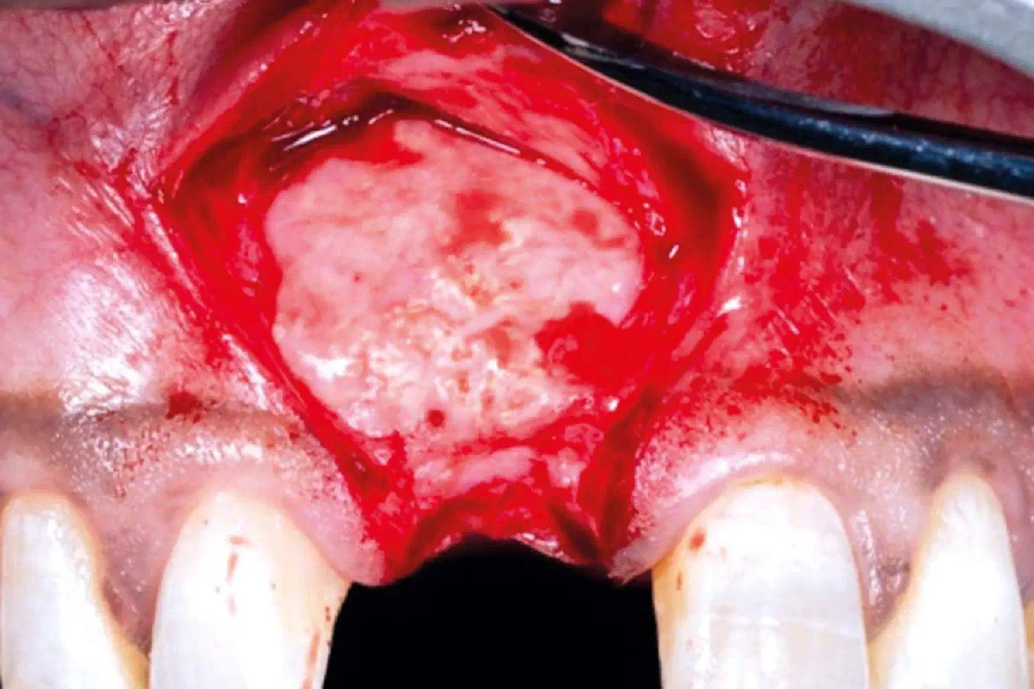
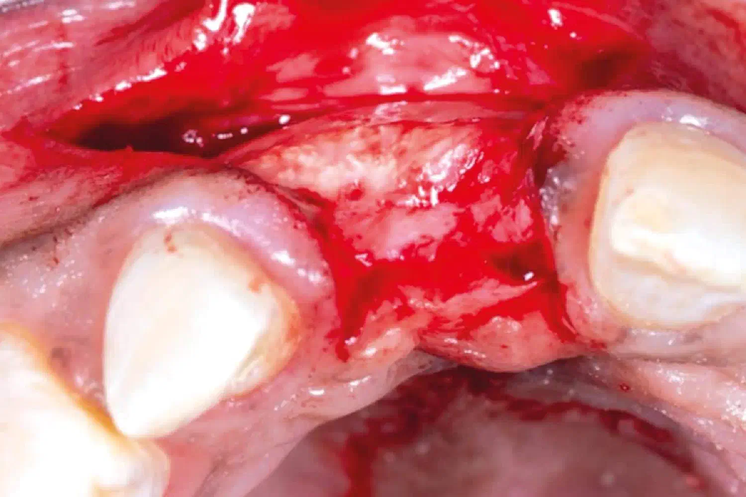
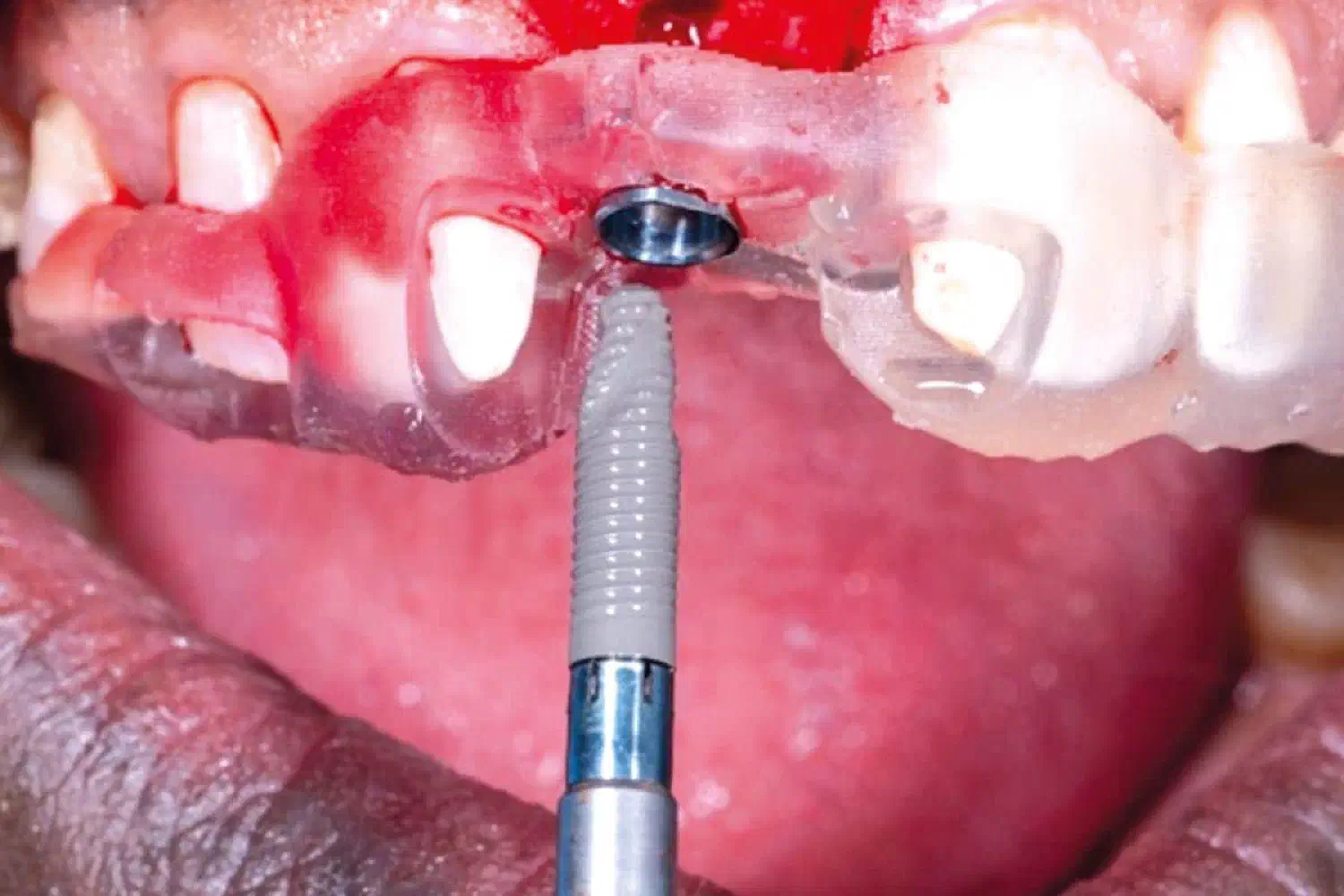
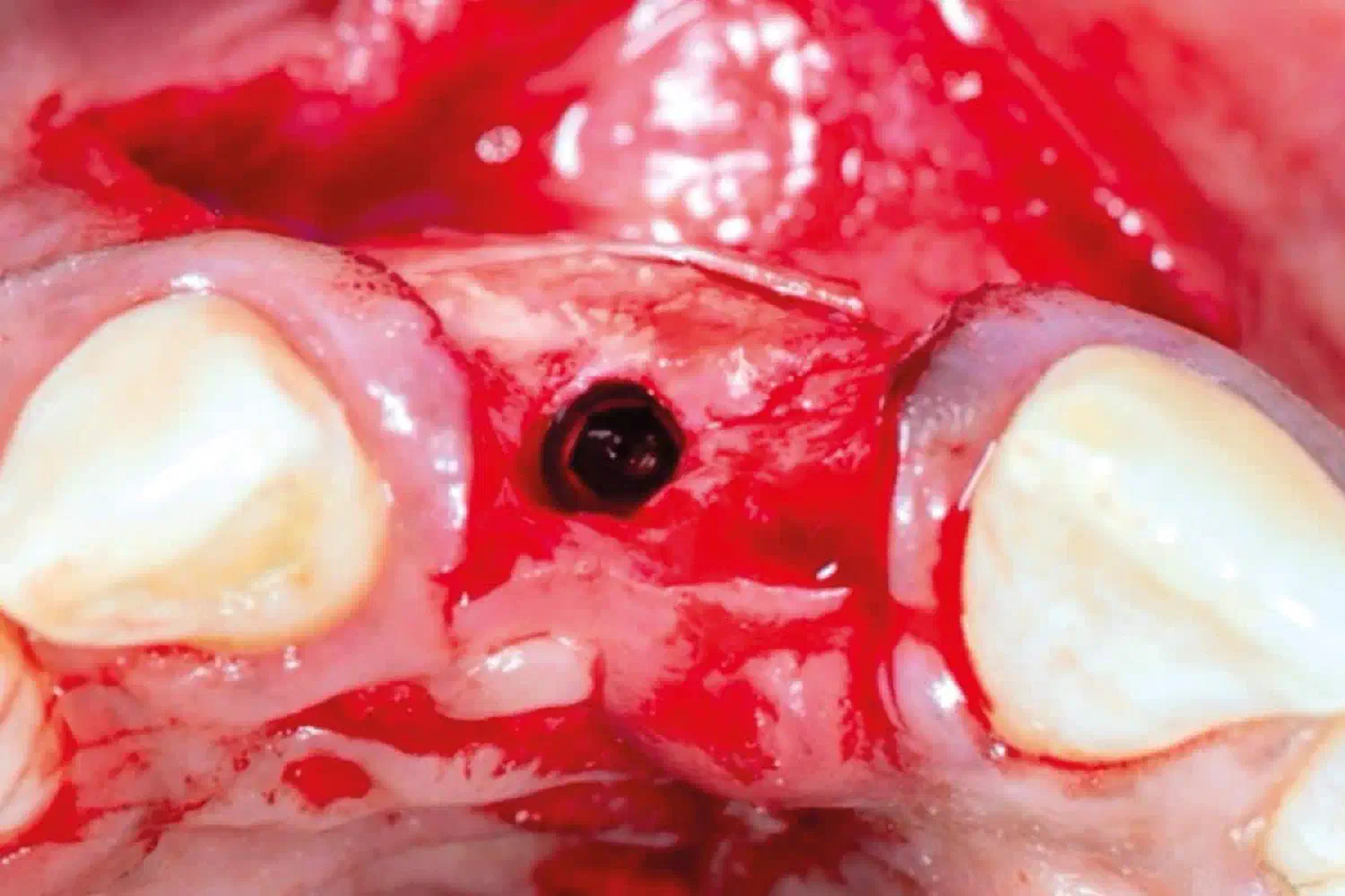
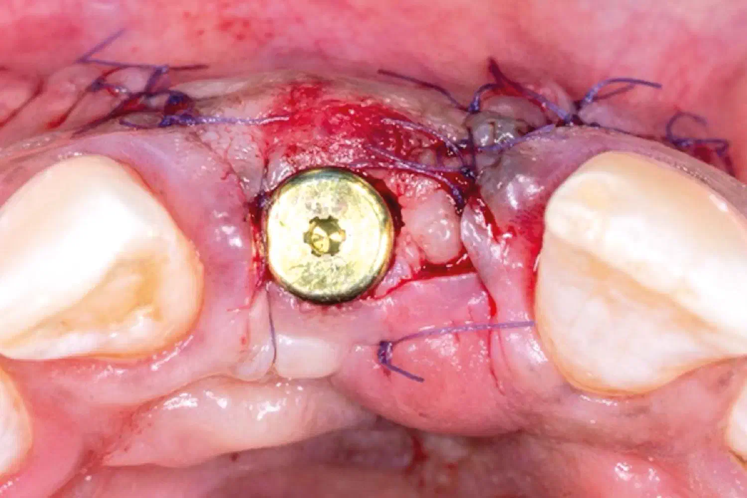
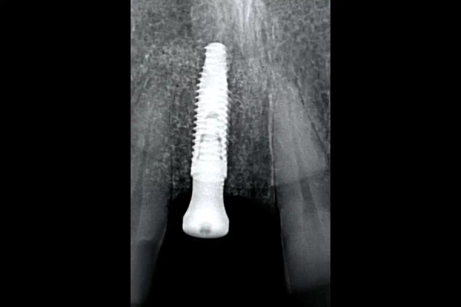
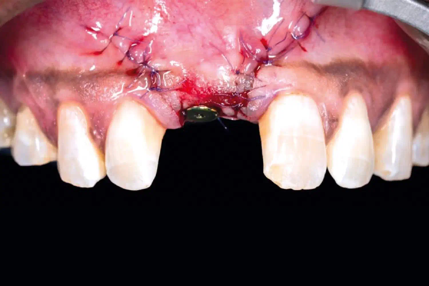
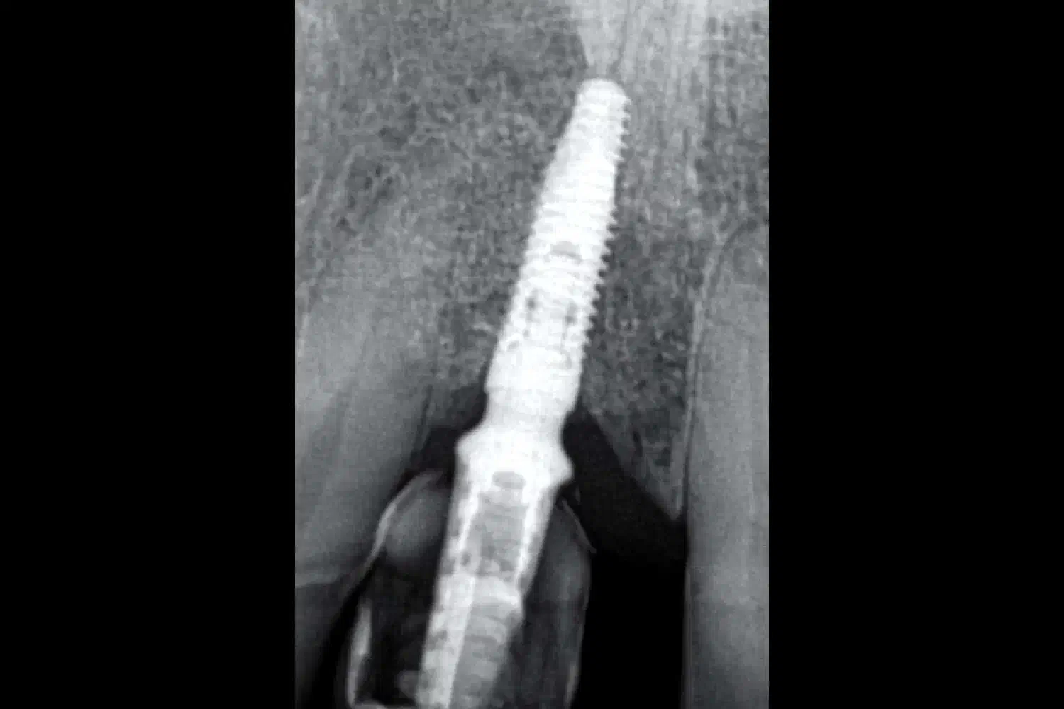
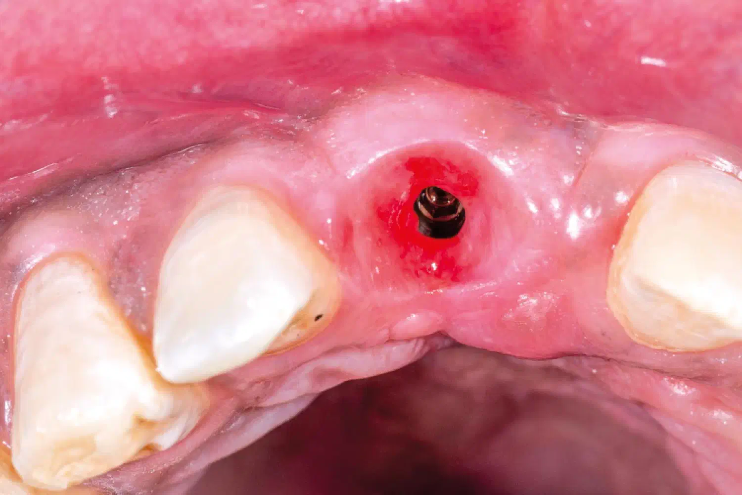
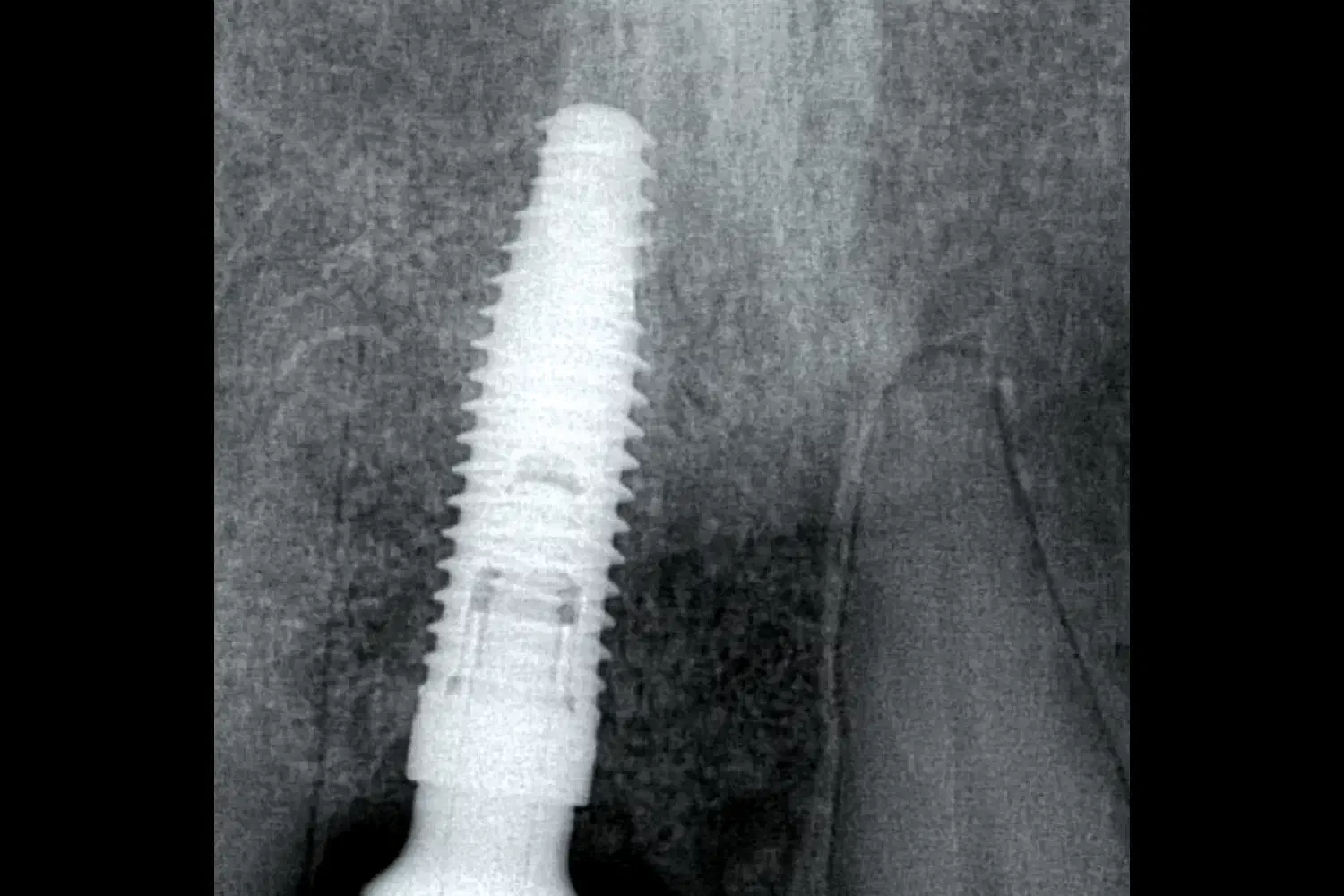
After 5 months of uneventful healing full thickness flap elevation was done to expose and remove the PTFE membrane and the titan pins . The cerabone® +Hya particles were integrated into the bone and a high quality and sufficient volume of regenerate bone was observed. An implant was placed following a fully guided protocol attaining an insertion torque. Relining of the buccal aspect of the regenerated bone was done using a thin layer of cerabone® +HyA which was covered using Jason® membrane . A healing collar was attached at the end of the implantation procedure.
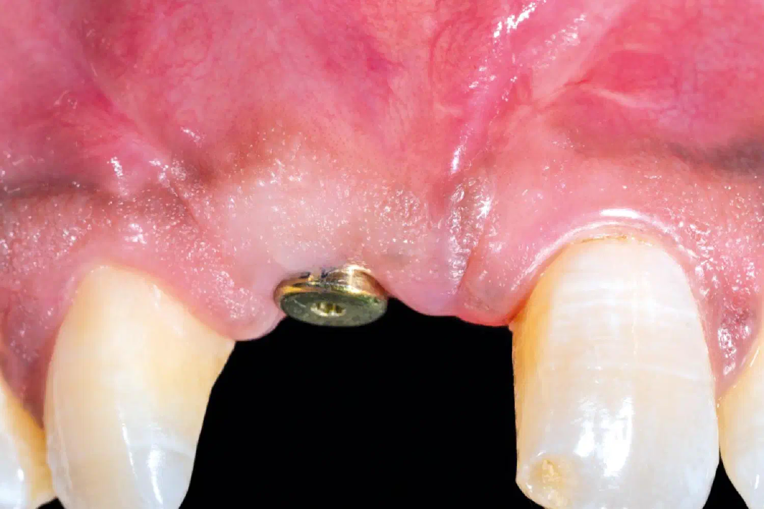
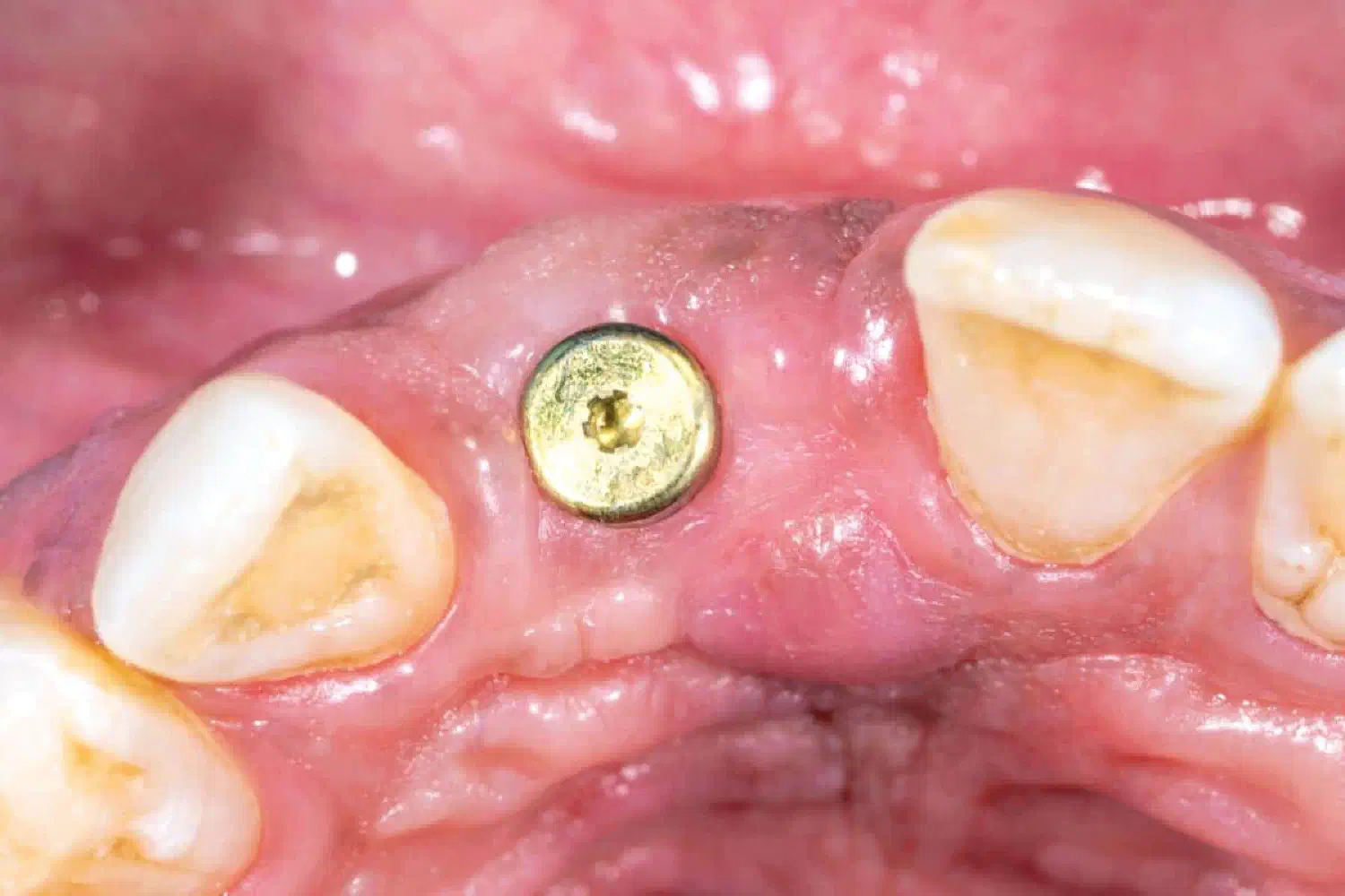
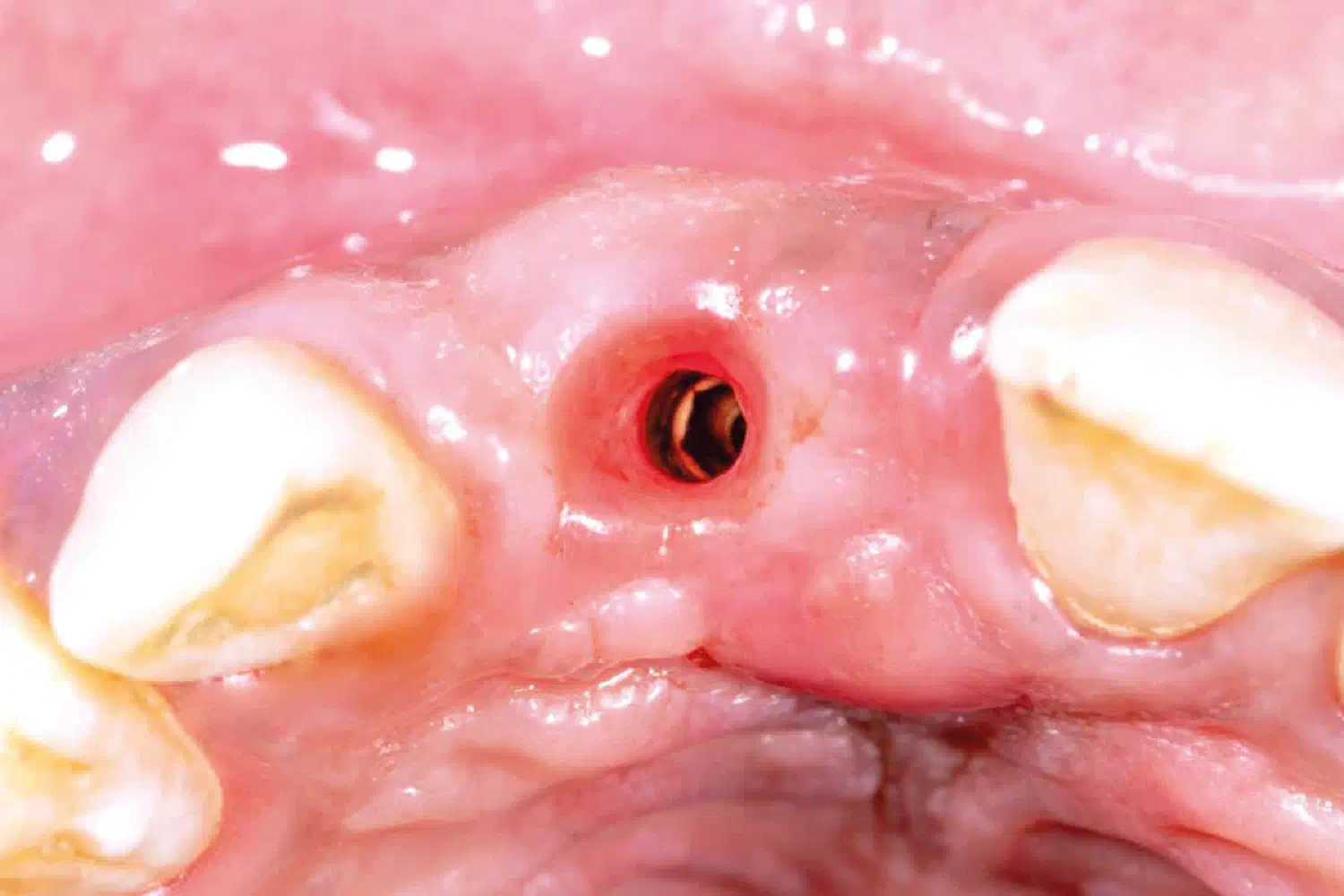
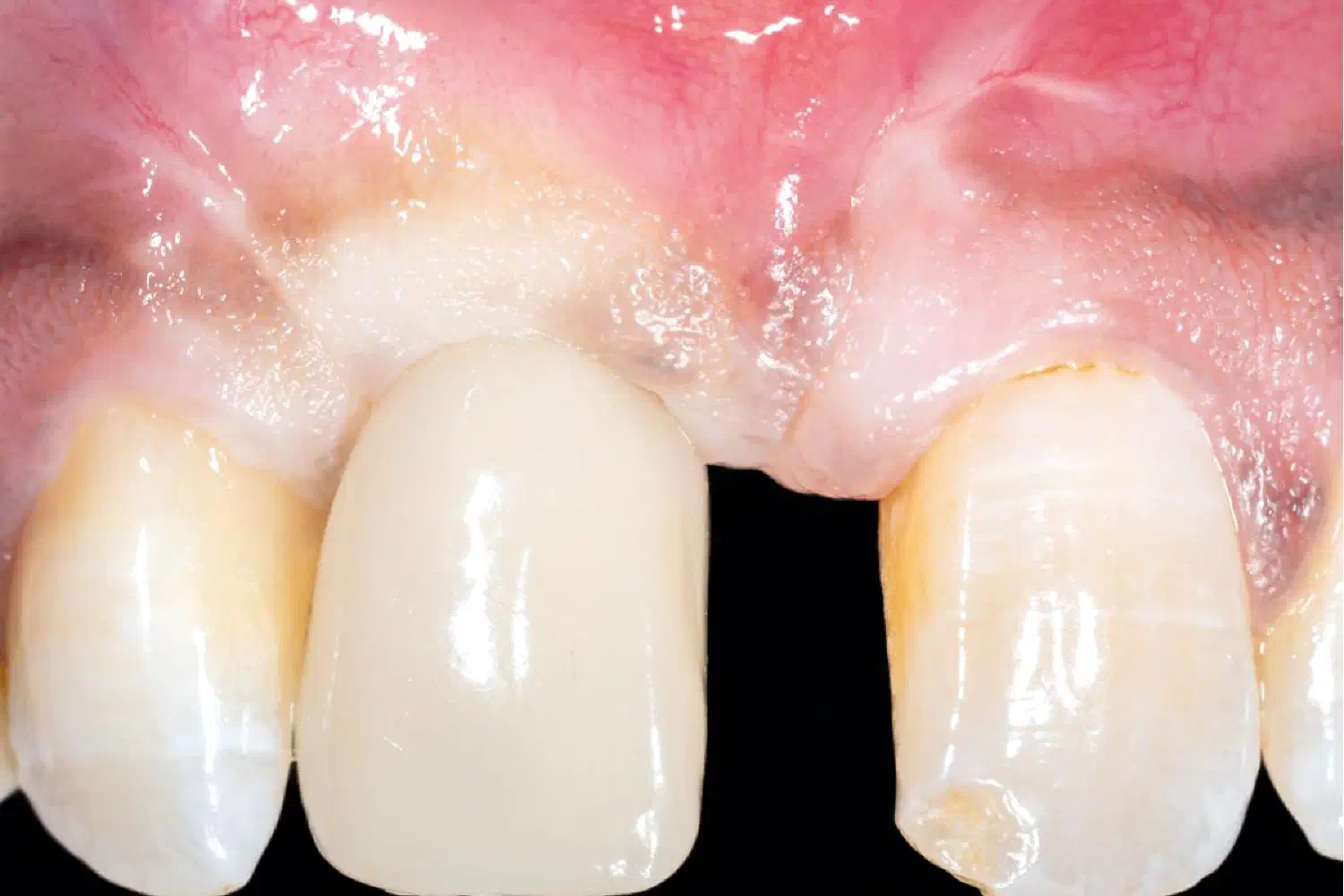
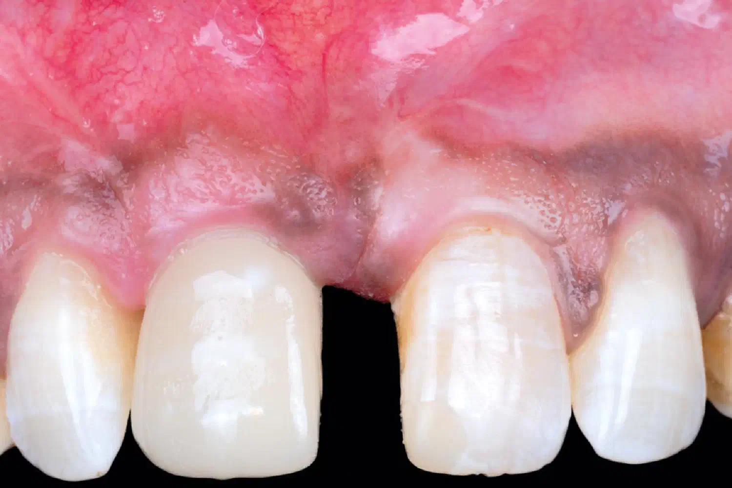
Following an uneventful osseintegration period of 8 weeks, soft tissue modulation with a provisional screw retained resin crown was commenced. This was carried out for a period of 4 months to achieve mucosal margin (level & scallop) and papilla heights similar to the left central incisor. A final restoration was delivered after no further soft tissue changes were observed and the patient was pleased with the aesthetic outcome.
The „Case Of The Month“ highlights every month a clinical case, which distinguished itself by the clinical results or the treatment concept in combination with the applied botiss biomaterials. The selection of the case is based on content relevance and quality of the documentation.