Acellular dermal collagen matrix

mucoderm®

  • Alternative to autologous soft tissue
  • Rapid revascularization
  • Complete remodelling into patient’s own tissue

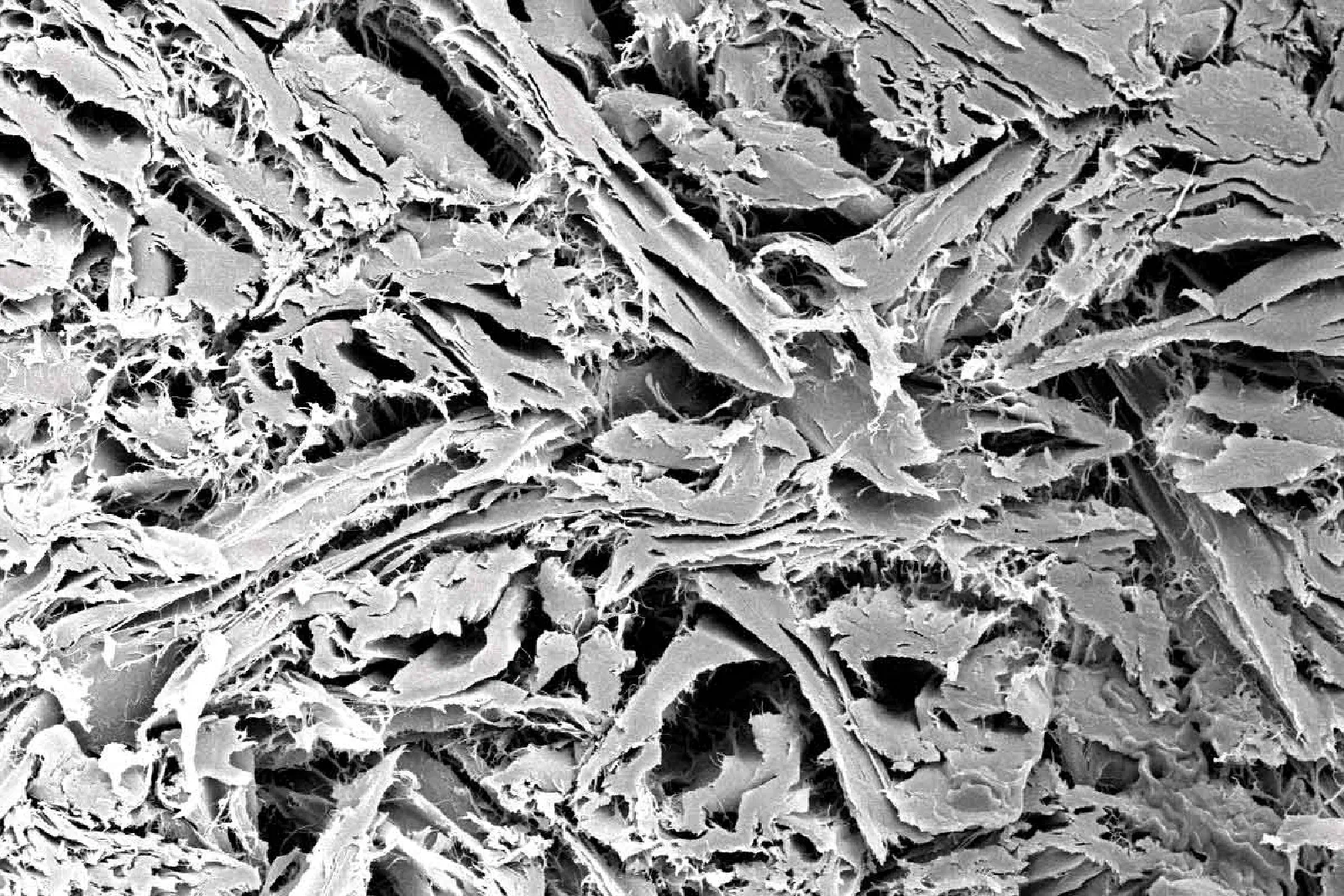
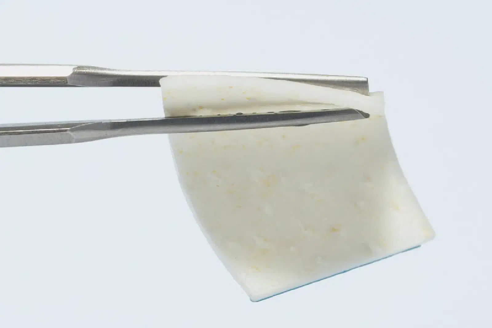
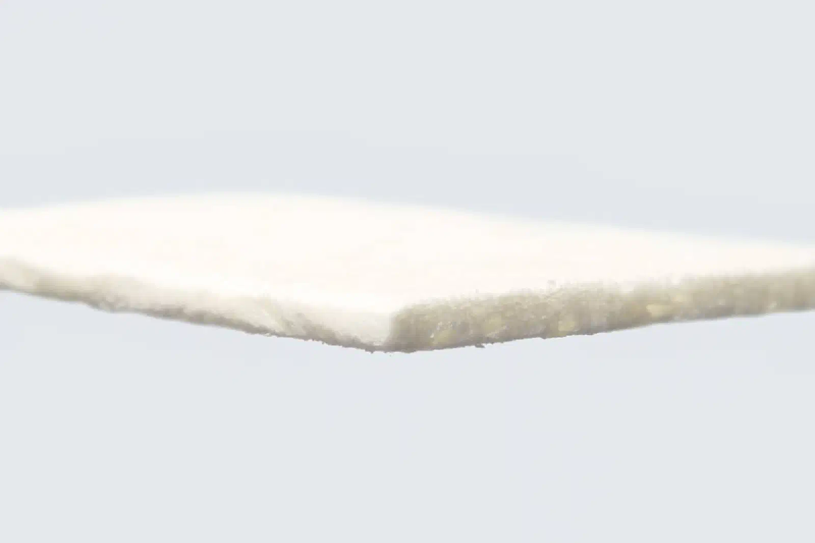
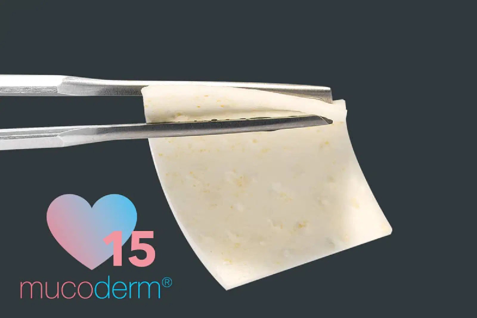
mucoderm® is an acellular collagen matrix that offers a safe alternative to autologous soft tissue transplants in a diverse range of soft tissue grafting indications. mucoderm® is derived from porcine dermis that undergoes a multi-step purification process, which removes all non-collagenous proteins and cells as well as potential immunogens, bacteria and viruses. The processing results in a three-dimensional, stable matrix, which consists of collagen type I and III with a natural collagen structure that resembles the human connective tissue¹,². After implantation mucoderm® is continuously remodeled into patients own soft tissue.
Indications · Handling tips · Specifications

Scaffold for ingrowing cells and vessels

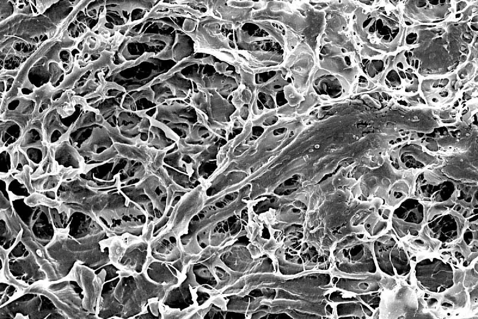
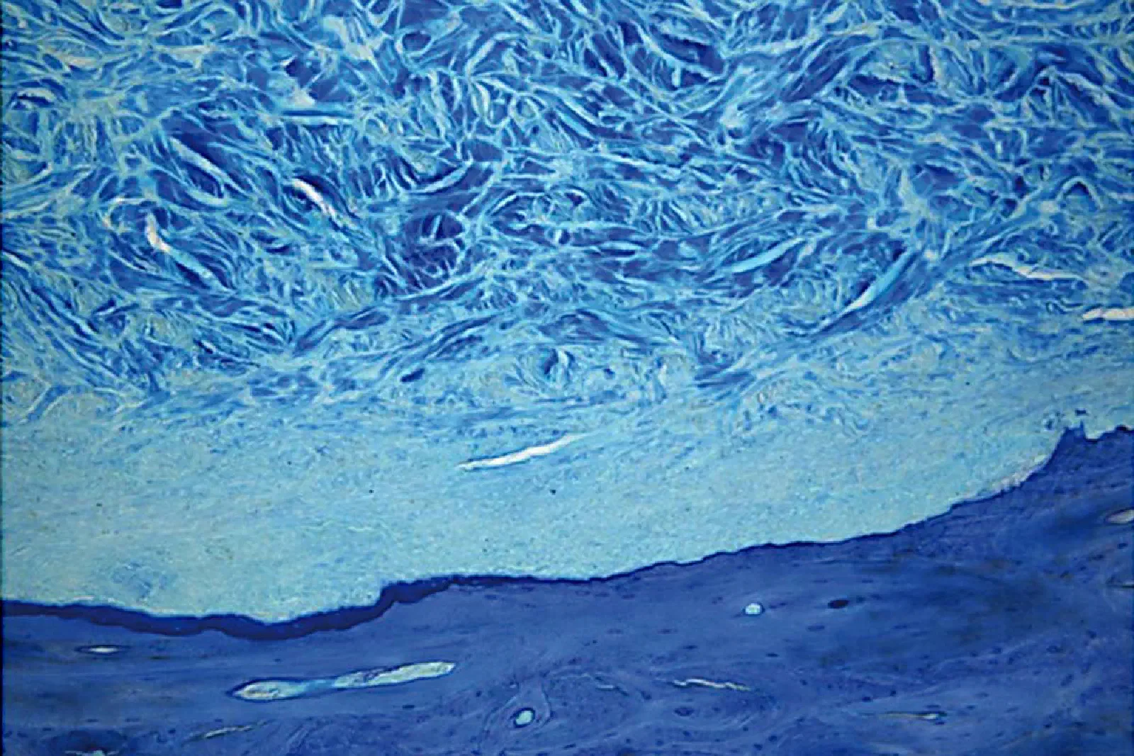
mucoderm® shows a high porosity and native collagen structure making it an excellent scaffold for ingrowing blood vessels and cells, thus supporting a fast revascularization and tissue integration³. Attracted by the signals of activated migrating and proliferating endothelial cells, blood vessels from the surrounding tissue will grow into the matrix. At the same time, fibroblasts adhere and spread onto the matrix. The simultaneous degradation of the matrix and the collagen production of adhering fibroblasts leads to a complete substitution of mucoderm® by the newly formed host tissue within about 6–9 months⁴⁻⁶.

Featured cases

Indications

mucoderm® can be applied as an alternative to autologous soft tissue transplants such as connective tissue graft (CTG) and free gingival graft (FGG) in various indications in periodontology and implantology. The use of mucoderm® is of particular advantage in situations where autologous transplants cannot be harvested in sufficient amount or quality (e.g. in thin biotype, shallow palate or covering of multiple recessions) or if patients are afraid of and do not agree with a tissue harvesting.

Implantology, periodontology and oral and CMF:

  • Soft tissue grafting in combination with GBR/GTR
  • Broadening of attached gingiva (instead of FGG)
  • Treatment of gingival recessions
  • Thickening of periimplant soft tissue
  • Closure of extraction sockets (socket seal technique)
  • Oral wound coverage after transplant harvesting or tumor surgery

Properties + advantages

  • Rapid revascularization and tissue integration
  • Soft tissue regeneration/augmentation without palatal autograft harvesting
  • Complete remodelling into patient’s own tissue in ~6–9 months
  • Can be easily applied and fixed by sutures
  • Can be cut into procedure-specific shape

Handling tips

Hydration

mucoderm® should always be applied after rehydration (in sterile saline, defect blood, or platelet concentrates). Rehydration should be performed for five to 20 minutes, depending on the technique used and the desired flexibility of the matrix. The flexibility of the mucoderm® increases with rehydration time. The rehydration protocol and its influence on the biomechanical properties of mucoderm® were analyzed in a study of Prof Dr. Adrian Kasaj⁸.

Trimming

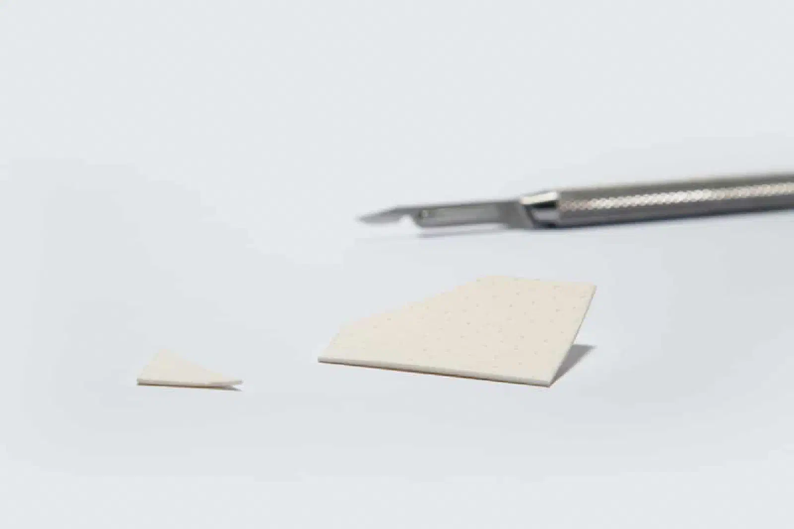
Size and shape of the matrix should be adapted to the size of the defect. After rehydration, mucoderm® can be easily trimmed to the desired size with a scalpel or scissors. Cutting or rounding the edges can prevent the perforation of the gingival tissue during the flap closure. For the coverage of multiple recessions, the surface of mucoderm® can be extended by cutting the matrix on alternating sides (mesh-graft technique) and pulling it.

Immobilization/fixation

Following application, mucoderm® should always be stabilized to avoid micro movements and ensure undisturbed revitalization, e.g. ingrowth of vessels and cells. When preparing a split flap, mucoderm® should be sutured to the intact periosteum to ensure close contact between the matrix and the periosteal wound bed. Single button or cross sutures may be used; the use of resorbable sutures is recommended. Both sides of the matrix are comparable, since mucoderm® shows a homogenous structure.

Exposure

mucoderm® can be left exposed for open healing in situations in which revitalization from the surrounding or underlying wound bed is ensured. Always avoid exposure of mucoderm® when used in recession coverage. A close flap is also recommended when simultaneously applied with bone grafting materials. Open healing is feasible in the case of a vestibuloplasty, if mucoderm® is sutured to the periosteum. In this case mucoderm® should be closely fixed to the periosteum. This facilitates an increase in the width of the attached gingiva but not in the thickening of the tissue. Open healing is also possible if only minor parts of the matrix are exposed, and revascularization is ensured by the surrounding margins of the flap or by the underlying periosteum. Please note that the degradation time depends on the extent of the exposure and will be faster due to bacterial decontamination and resorption.

Flap preparation

mucoderm® can be used in combination with all mucogingival surgical techniques, including coronally advanced flap and tunnel techniques. Notably, the classical coronally advanced flap or the modified coronally advanced flap ensure a good view on the prepared donor bed and facilitate the coronal repositioning of the flap over the matrix. Advanced flaps need to be sufficiently mobilized to avoid tension of the soft tissue. A proper vascular supply from the prepared flap is critical to achieve an appropriate revascularization of the mucoderm® matrix⁹. In particular, split flaps must be sufficiently thick to ensure revitalization of the matrix and the integration into the patient’s own connective tissue. When applying mucoderm® for recession coverage, special attention must be paid to achieve sufficient flap mobilization and tension-free closure.

Combination with Straumann® Emdogain®

Straumann® Emdogain® is an easy-to-apply gel containing enamel matrix derivative originating from non-erupted porcine tooth buds. As a component of embryonic tissues it is designed to promote predictable regeneration of hard and soft tissue due to periodontal disease or trauma. Gingival recessions may be treated using mucoderm® and Straumann® Emdogain® together in a single approach. Always make sure that Straumann® Emdogain® is applied all over the root surfaces before the mucoderm® matrix is placed over the roots.

Specific facts

Highest safety

The particular, certified multi-stage cleaning process of mucoderm® effectively removes all non-collagenous proteins and cells as well as potential immunogens, bacteria and viruses. Hence, mucoderm® is an absolutely safe and pure collagen type I and III matrix. mucoderm® is a medical device regulated according to EC-Guidelines. Manufacturing of mucoderm® is subject to a quality control system based on international standards (e.g. EN ISO 13485), and is regularly audited by the notified body and authorities.

Native collagen structure

mucoderm® is a native collagen matrix, meaning that the natural properties of the original tissue (dermis) are preserved during the production process. This is the basis for its superior handling properties such as tear resistance and dimensional stability. Preservation of the natural structure is also the reason that mucoderm® structure strongly resembles the architecture of the human dermis¹, making the matrix a suitable alternative to autologous gingival transplants.

Tissue integration, revascularization and degradation

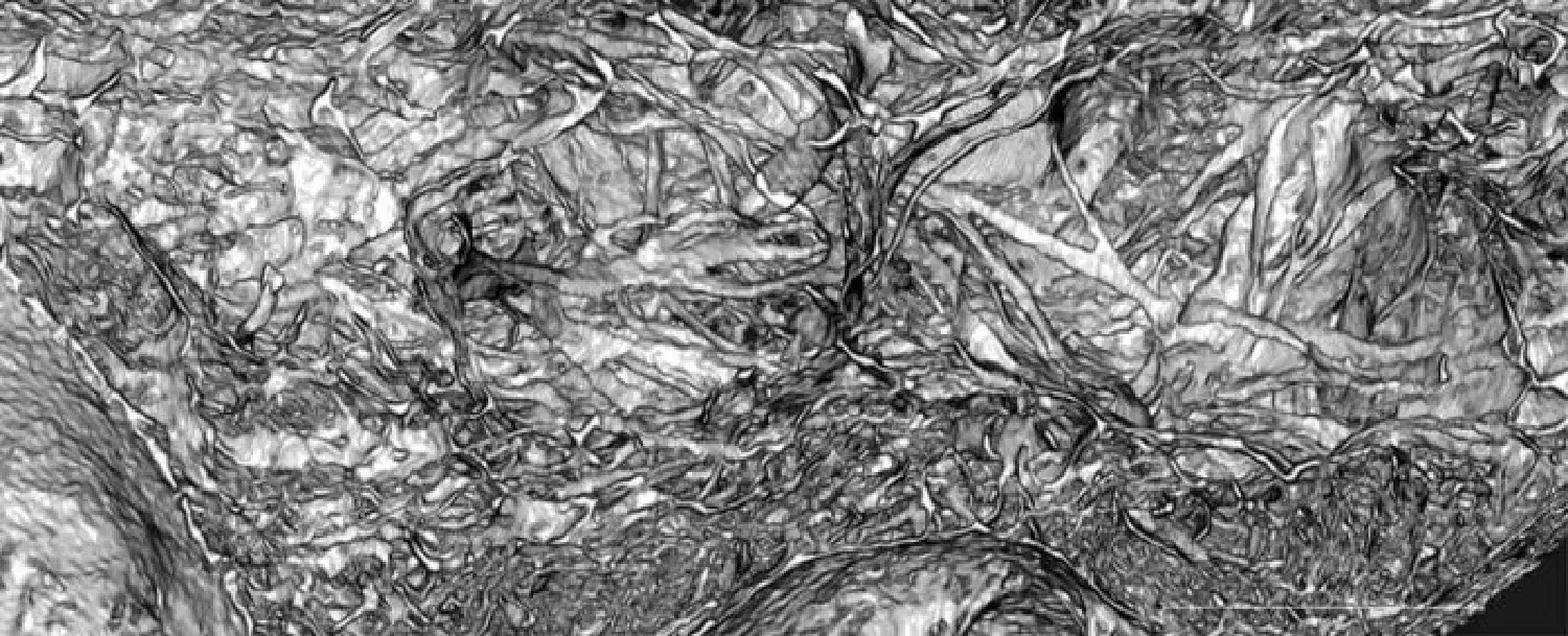
mucoderm® is characterized by a porous collagen structure, visualized by the innovative synchrotron-based X-ray tomography⁷: Synchrotron-based X-ray tomographic microscopy for visualization of three-dimensional collagen matrices.

Synchrotron-based x-ray image showing the porous structure of mucoderm®

Synchrotron-based x-ray image showing the porous structure of mucoderm®

This unique structure makes mucoderm® an ideal scaffold for ingrowth of blood vessels and cells and promotes fast tissue integration and revascularization. Tissue integration and degradation were analyzed in an animal (rat model) study of Prof. Daniel Rothamel⁴. After only two weeks, mucoderm® shows an extensive ingrowth of blood vessels as well as an inflammation-free healing with superficial cell invasion. In the following four to eight weeks, a continuous degradation with increasing homogeneous cell distribution can be observed. After eight weeks, still 20% of the original matrix volume is available as a scaffold for the formation and reorganization of connective tissue. After twelve weeks, mucoderm® is almost completely replaced by newly formed connective tissue.

Combination of mucoderm® with enamel matrix proteins

mucoderm® supports the maintenance and thickening of gingival tissue, which can be particularly advantageous in thin biotypes. The additional application of Straumann® Emdogain® during recession coverage with mucoderm® can:

  • improve the qualitative attachment¹⁰,¹¹;
  • stimulate angiogenesis¹²,¹³ and thus improve the revascularization and integration of mucoderm®;
  • increase the quantity of keratinized tissue, which can be beneficial in case of lack or absence of keratinized gingiva¹⁴.

References

  1. Ramachandra SS, Rana R, Reetika S, Jithendra KD. Options to avoid the surgical site: a review of literature. Cell Tissue Bank 2014, 15(3): 297-305.
  2. Meyer et al. 1982. Collagen fiber arrangement in the skin of the pig. J Anat.;134:139-48.
  3. Pabst AM, Happe A, Callaway A, Ziebart T, Stratul SI, Ackermann M, Konerding MA, Willershausen B, Kasaj A. In vitro and in vivo characterization of porcine acellular dermal matrix for gingival augmentation procedures. Periodont Res. 2014, 49(3): 37-81
  4. Rothamel et al. 2014. Biodegradation pattern and tissue integration of native and cross-linked porcine collagen soft tissue augmentation matrices – an experimental study in the rat. Head and Face 27;10:10.
  5. Puisys et al. 2019. Clinical and Histologic Evaluations of Porcine-Derived Collagen Matrix Membrane Used for Vertical Soft Tissue Augmentation: A Case Series. Int J Periodontics Restorative Dent. 39(3):341-347.
  6. Zafiropoulos et al. 2016. Changes of the peri-implant soft tissue thickness after grafting with a collagen matrix. J Indian Soc Periodontol.; 20(4):441-445.
  7. Pabst AM, Wagner W, Kasaj A, Gebhardt S, Ackermann M, Astolfo A, Marone F, Haberthür D, Enzmann F, Konerding M A. Synchrotronbased X-ray tomographic microscopy for visualization of three-dimensional collagen matrices. Clin Oral Investig 2015, 19(2):561-4.
  8. Kasaj A, Levin L, Stratul SI, Götz H, Schlee M, Rütters CB, Konerding MA, Ackermann M, Willershausen B, Pabst AM. The influence of various rehydration protocols on biomechanical properties of different acellular tissue matrices. Clin Oral Invest. 2015.
  9. Mörmann W, Ciancio SG. Blood supply of human gingiva following periodontal surgery. A fluorescein angiographic study. J Periodontol. 1977 Nov;48(11):681-92
  10. McGuire MK, Cochran DL. Evaluation of human recession defects treated with coronally advanced flaps and either enamel matrix derivative or connective tissue. Part 2: Histological evaluation. J Periodontol. 2003 Aug;74(8):1126-35.
  11. Shirakata Y, Sculean A, Shinohara Y, Sena K, Takeuchi N, Bosshardt DD, Noguchi K. Healing of localized gingival recessions treated with a coronally advanced flap alone or combined with an enamel matrix derivative and a porcine acellular dermal matrix: a preclinical study. Clin Oral Investig. 2015 Nov 27.
  12. Kasaj A, Meister J, Lehmann K, Stratul SI, Schlee M, Stein JM, Willershausen B, Schmidt M. The influence of enamel matrix derivative on the angiogenic activity of primary endothelial cells. J Periodontal Res. 2012 Aug;47(4):479-87.
  13. Aspriello SD, Zizzi A, Spazzafumo L, Rubini C, Lorenzi T, Marzioni D, Bullon P, Piemontese M. Effects of enamel matrix derivative on vascular endothelial growth factor expression and microvessel density in gingival tissues of periodontal pocket: a comparative study. J Periodontol. 2011 Apr;82(4):606-12.
  14. Pilloni A, Paolantonio M, Camargo PM. Root coverage with a coronally positioned flap used in combination with enamel matrix derivative: 18-month clinical evaluation. J Periodontol. 2006 Dec;77(12):2031-9.
botiss worldwide

botiss
biomaterials
Benelux

Schijfstraat 24
5061 KB Oisterwijk
The Netherlands

Available on weekdays
from 08:30 to 17:00

Shop shopnow News botissnews